
A subscription to JoVE is required to view this content. Sign in or start your free trial.
Method Article
Stochastic Noise Application for the Assessment of Medial Vestibular Nucleus Neuron Sensitivity In Vitro
In This Article
Summary
Galvanic vestibular stimulation in humans exhibits improvements in vestibular function. However, it is unknown how these effects occur. Here, we describe how to apply sinusoidal and stochastic electrical noise and evaluate appropriate stimulus amplitudes in individual medial vestibular nucleus neurons in the C57BL/6 mouse.
Abstract
Galvanic vestibular stimulation (GVS) has been shown to improve balance measures in individuals with balance or vestibular impairments. This is proposed to be due to the stochastic resonance (SR) phenomenon, which is defined as application of a low-level/subthreshold stimulus to a non-linear system to increase detection of weaker signals. However, it is still unknown how SR exhibits its positive effects on human balance. This is one of the first demonstrations of the effects of sinusoidal and stochastic noise on individual neurons. Using whole-cell patch clamp electrophysiology, sinusoidal and stochastic noise can be applied directly to individual neurons in the medial vestibular nucleus (MVN) of C57BL/6 mice. Here we demonstrate how to determine the threshold of MVN neurons in order to ensure the sinusoidal and stochastic stimuli are subthreshold and from this, determine the effects that each type of noise has on MVN neuronal gain. We show that subthreshold sinusoidal and stochastic noise can modulate the sensitivity of individual neurons in the MVN without affecting basal firing rates.
Introduction
The vestibular (or balance) system controls our sense of balance by integrating auditory, proprioceptive, somatosensory and visual information. Degradation of the vestibular system has been shown to occur as a function of age and can result in balance deficits1,2. However, therapies targeting the functioning of the vestibular system are scarce.
Galvanic Vestibular Stimulation (GVS) has been shown to improve balance measures, autonomic functioning and other sensory modalities within humans3,4,5,6. These improvements are said to be due to the Stochastic Resonance (SR) phenomenon, which is the increase in the detection of weaker signals in non-linear systems via the application of subthreshold noise7,8. These studies have shown improvements in static9,10 and dynamic11,12 balance, and vestibular output tests such as Ocular Counter Roll (OCR)13. However, many of these studies have used different combinations of stimulus parameters such as white noise9, colored noise13, different stimulus frequency ranges and thresholding techniques. Therefore, optimal stimulus parameters remain unknown and this protocol can assist with determining the most effective parameters. Besides stimulus parameters, the type of stimulus is also important in therapeutic and experimental efficacy. The above work in humans was performed using electrical noise stimuli, whilst much of the in vivo animal work has used mechanical14,15 or optogenetic16 noise stimuli. This protocol will use electrical noise to examine the effects on vestibular nuclei.
Previously, application of GVS to stimulate primary vestibular afferents was been performed in vivo in squirrel monkeys17, chinchillas18, chicken embryos15 and guinea pigs14. However, only two of these studies examined the effect GVS has on the gain of primary vestibular afferents14,15. These experiments were performed in vivo meaning that the precise patterns of stimulation imposed on vestibular nuclei cannot be determined. To our knowledge, only one other study has applied stochastic noise to individual enzymatically dissociated neurons in the central nervous system19. However, no experiments have been performed in the central vestibular nuclei to assess appropriate stimulus parameters and thresholding techniques, making this protocol more precise in determining stimulus effects on individual neurons within the vestibular nuclei.
Here, we describe how to apply sinusoidal and stochastic (electrical) noise directly to individual neurons in the medial vestibular nucleus (MVN), determine neuronal threshold and measure changes in gain/sensitivity.
Access restricted. Please log in or start a trial to view this content.
Protocol
All experimental protocols described were approved by the University of Sydney Animal Ethics Committee (approved protocol number: 2018/1308).
1. Animals
NOTE: Mice were obtained from the Australian Rodent Centre (ARC; Perth, Australia) and held at the Medical Foundation Building Animal Facility at the University of Sydney.
- Maintain the mice on a normal 12 h light/dark cycle with environmental enrichment.
- Use male and female C57BL/6 mice (3–5 weeks old) for all experiments.
2. Preparation of Solutions
- Prepare 1 L of artificial cerebrospinal fluid (ACSF) composed of 29 mM NaHCO3, 11 mM glucose, 120 mM NaCl, 3.3 mM KCl, 1.4 mM NaH2PO4, 2.2 mM MgCl2, 2.77 mM CaCl2.
- Prepare 200 mL of sucrose-ACSF (sACSF) containing 29 mM NaHCO3, 11 mM glucose, 241.5 mM sucrose, 3.3 mM KCl, 1.4 mM NaH2PO4, 2.2 mM MgCl2, 2.77 mM CaCl2. Prior to the inclusion of CaCl2 to the ACSF and sACSF, gas the solutions with carbogen (95 % O2 and 5 % CO2) to establish a pH of 7.4 and avoid calcium precipitation (cloudiness).
- Prepare K+-based intracellular solution composed of 70 mM potassium gluconate, 70 mM KCl, 2 mM NaCl, 10 mM HEPES, 4 mM EGTA, 4 mM Mg2-ATP, 0.3 mM Na3-GTP; with a final pH of 7.3 (adjusted using KOH).
NOTE: It is recommended to filter intracellular solutions with 0.22 µm filters and store 0.5 mL aliquots of the solution at -20 °C.
3. Preparation of the Brainstem
- Prior to brainstem extraction, equilibrate the sACSF with carbogen and cool at -80 °C for 25 min so that an ice slurry is formed.
- Anaesthetize the mouse with isoflurane (3–5 %) saturated in oxygen (3 mL/min). Once the hind paw reflexes are absent, decapitate the mouse with sharp stainless-steel scissors.
- Expose the skull by making a sagittal incision in the skin using a razor blade (#22 rounded).
- Using the pointed end of a pair of standard pattern scissors make a small incision at the lambda and cut along the longitudinal fissure.
- Carefully reflect away the paired parietal bones and the occipital bones using a pair of shallow-bend Pearson rongeurs.
NOTE: During this whole procedure the brain is continuously bathed in situ using the previously prepared ice-cold sACSF slurry. - Isolate the brainstem from the forebrain and its bony encasing using a razor blade (#11 straight) to cut down the parieto-occipital sulcus and at the caudal medulla.
- Mount the isolated brainstem ventral end down on a previously cut trapezoidal polystyrene block. Remove excess fluid around the dissected tissue with a wick of tissue paper to ensure good tissue adhesion to the cutting stage.
NOTE: The polystyrene block is cut in a trapezoidal shape, to ensure the rostral end of the midbrain fits and tapers into the spinal cord. - Use cyanoacrylate glue to fix the polystyrene block with the attached brainstem rostral end down to the cutting stage.
- Using an advance speed of 0.16 mm/s and vibration amplitude of 3.00 mm, prepare 200 µm transverse slices of the MVN.
NOTE: Location of the MVN is determined using the Paxinos and Franklin mouse brain atlas (Figures 79–89)20. The MVN (listed as MVe in atlas) lies immediately ventrolateral to the 4th ventricle and is largest right before the attachment of the cerebellum (between the inferior colliculi and the obex). - Use a plastic-trimmed pipette to transfer slices onto a filter paper disc sitting in carbogenated ACSF at 25 °C for at least 30 min prior to recording.
4. Instruments
- Use a standard electrophysiological setup to perform whole-cell patch clamp techniques21.
- Prepare micropipettes using a two-step protocol (heat step 1: 70; heat step 2: 45) on a micropipette puller (see the Table of Materials). Micropipettes should have a final resistance ranging 3–5 MΩ with internal solution when placed in the bath.
NOTE: Settings used may vary depending on the temperature within room and can change quite frequently.
5. Whole-cell Patch Clamp Electrophysiology
- To obtain whole-cell patch clamp recordings from individual neurons in the MVN, a K+-based internal solution is used within the recording pipette.
- Transfer a single tissue slice from the incubation chamber to the recording chamber and secure the slice using a nylon thread on a U-shaped weight. Continuously perfuse the recording chamber with carbogenated-ACSF at 25 °C at a flow rate of 3 mL/min.
- After filling a micropipette with internal solution, locate the MVN using a low power (10x) objective lens. Using a high-power (40x) objective, individual neurons within the MVN can be located.
NOTE: Cell quality is essential in ensuring quality recordings and durability of the cell when attempting to achieve the whole-cell configuration. A good cell will demonstrate spherical shape, a reflective cell membrane and an invisible nucleus. A bad cell will have a large visible nucleus (egg-like) and a swollen/shrunken appearance. - Before breaching the tissue with the pipette, apply a small amount of positive pressure to push debris away from the pipette tip.
- Move the pipette using the micromanipulator towards the chosen neuron and a small dimple should form on the neuronal membrane. Release positive pressure and apply a small amount of negative pressure.
- Once a 1 GΩ seal is achieved, apply gentle short and sharp positive pressure to the pipette holder through the suction port to rupture the membrane and create a whole-cell configuration.
- Make whole-cell current clamp recordings using standard techniques21,22.
6. Applying Sinusoidal and Stochastic Noise to Individual Medial Vestibular Nucleus Neurons
- Apply the stochastic and sinusoidal noise at a range of amplitudes from 3 to 24 pA to determine neuronal threshold and firing rate.
- Determine the sensory threshold by grouping lower and higher stimulus intensities and perform an ANOVA to observe any differences (as shown in Supplementary Figure 1).
- Calculate the average firing rate over the 10 s period where the depolarizing current step was/will be injected for each individual current level (i.e., 7 total episodes; Figure 1).
- Use the average firing rate values to generate a firing rate versus current plot and perform a linear regression analysis to determine the gradient of the line of best fit. The gradient of the line of best fit is indicative of the neuronal gain22.

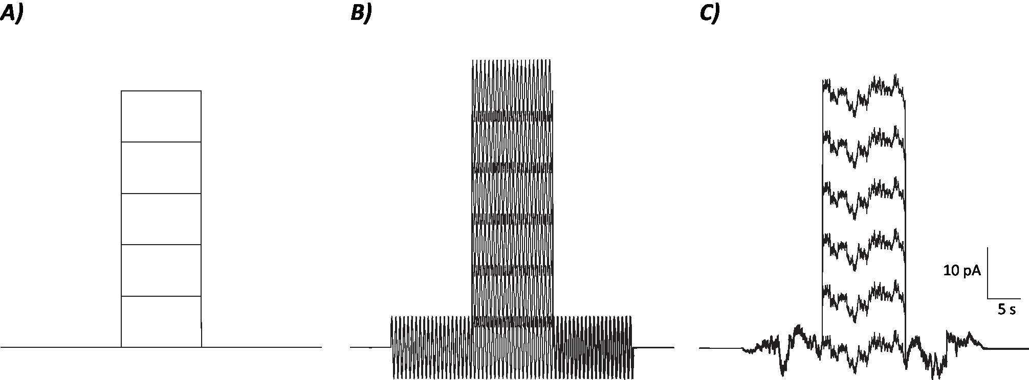
Figure 1: Diagrammatic profiles of control, sinusoidal and stochastic noise protocols. (A) Control (no noise) protocols applied to MVN neurons. (B) Sinusoidal noise protocol with a frequency of 2 Hz. (C) Stochastic noise protocols where majority of the power spectrum is ≤2 Hz. Each protocol presented here has an amplitude of ±6 pA with a 10 s depolarizing current increasing by 10 pA up to 50 pA. The true stimulus does not have a depolarizing current step and is therefore the first episode of these protocols to determine neuronal gain changes. Please click here to view a larger version of this figure.
Access restricted. Please log in or start a trial to view this content.
Results
Initial recordings can provide information about the effects that sinusoidal and stochastic noise have on basal firing rates of individual MVN neurons and how the stimuli effect the gain of neurons. Figure 2 shows that neither sinusoidal nor stochastic noise change basal firing rates of MVN neurons when compared to control (no noise) recordings. This information is crucial for determining the threshold of the individual neurons. During the application of galvanic vestibul...
Access restricted. Please log in or start a trial to view this content.
Discussion
The effects of galvanic vestibular stimulation (GVS) on the vestibular system has been highlighted in vivo in humans3,13,23, guinea pigs14, rodents18 and non-human primates24. However, none of these studies have assessed the direct impact of electrical noise on the sensitivity of individual neurons in the vestibular system. Here we demonstrate the first in v...
Access restricted. Please log in or start a trial to view this content.
Disclosures
The authors declare no conflicts of interest.
Acknowledgements
SPS was supported by the University of Sydney postgraduate research scholarship.
Access restricted. Please log in or start a trial to view this content.
Materials
| Name | Company | Catalog Number | Comments |
| CaCl | Scharlau | CA01951000 | Used for ACSF and sACSF |
| D-(+)-Glucose | Sigma | G8270 | Used for ACSF and sACSF |
| EGTA | Sigma | E0396-25G | Used for K-based intracellular solution |
| HEPES | Sigma | H3375-25G | Used for K-based intracellular solution |
| KCl | Chem-supply | PA054-500G | Used for ACSF, sACSF and intracellular solution |
| K-gluconate | Sigma | P1847-100G | Used for K-based intracellular solution |
| Mg-ATP | Sigma | A9187-500MG | Used for K-based intracellular solution |
| MgCl | Chem-supply | MA00360500 | Used for ACSF and sACSF |
| Na3-GTP | Sigma | G8877-100MG | Used for K-based intracellular solution |
| NaCl | Chem-supply | SO02270500 | Use for ACSF and intracellular solution |
| NaH2PO4•2H2O | Ajax | AJA471-500G | Used for ACSF and sACSF |
| NaHCO3 | Sigma | S5761-1KG | Used for ACSF and sACSF |
| Sucrose | Chem-supply | SA030-500G | Used for sACSF |
| Isoflurane | Henry Schein | 1169567762 | Used for anaesthetising mice |
| EQUIPMENT | |||
| Borosilicate glass capillaries | Warner instruments | GC150T-7.5 | 1.5 mm OD, 1.16 mm ID, 7.5 cm length |
| Data acquisition software | Axograph | Used for electrophysiology and analysis | |
| Friedmen-Pearson Rongeurs | World precision instruments | 14089 | Used for dissection |
| Micropipette puller | Narishige | PP-830 | Used for micropipette |
| Multiclamp amplifier | Axon instruments | 700B | Used for electrophysiology |
| pH meter | Sper scientific | 860033 | Used for internal solution |
| Standard pattern scissors | FST | 14028-10 | Used for dissection |
| Sutter micromanipulator | Sutter | MP-225/M | Used for electrophysiology |
| Upright microscope | Olympus | BX51WI | Used for electrophysiology |
| Vibratome | Leica | VT1200 | Used for slicing brain tissue |
References
- Amiridis, I. G., Hatzitaki, V., Arabatzi, F. Age-induced modifications of static postural control in humans. Neuroscience Letters. 350 (3), 137-140 (2003).
- Iwasaki, S., Yamasoba, T. Dizziness and imbalance in the elderly: age-related decline in the vestibular system. Aging and disease. 6 (1), (2015).
- Fujimoto, C., et al. Noisy galvanic vestibular stimulation induces a sustained improvement in body balance in elderly adults. Scientific Reports. 6, 37575(2016).
- Breen, P. P., et al. Peripheral tactile sensory perception of older adults improved using subsensory electrical noise stimulation. Medical Engineering & Physics. 38 (8), 822-825 (2016).
- Yamamoto, Y., Struzik, Z. R., Soma, R., Ohashi, K., Kwak, S. Noisy vestibular stimulation improves autonomic and motor responsiveness in central neurodegenerative disorders. Annals of Neurology. 58 (2), 175-181 (2005).
- Soma, R., Nozaki, D., Kwak, S., Yamamoto, Y. 1/f noise outperforms white noise in sensitizing baroreflex function in the human brain. Physical Review Letters. 91 (7), 078101(2003).
- Wiesenfeld, K., Moss, F. Stochastic resonance and the benefits of noise: from ice ages to crayfish and SQUIDs. Nature. 373 (6509), 33-36 (1995).
- Moss, F., Ward, L. M., Sannita, W. G. Stochastic resonance and sensory information processing: a tutorial and review of application. Clinical Neurophysiology. 115 (2), 267-281 (2004).
- Goel, R., et al. Using low levels of stochastic vestibular stimulation to improve balance function. PloS one. 10 (8), e0136335(2015).
- Inukai, Y., et al. Effect of noisy galvanic vestibular stimulation on center of pressure sway of static standing posture. Brain Stimulation: Basic, Translational, and Clinical Research in Neuromodulation. 11 (1), 85-93 (2018).
- Mulavara, A. P., et al. Using low levels of stochastic vestibular stimulation to improve locomotor stability. Frontiers in Systems Neuroscience. 9, 117(2015).
- Iwasaki, S., et al. Noisy vestibular stimulation increases gait speed in normals and in bilateral vestibulopathy. Brain stimulation. 11 (4), 709-715 (2018).
- Serrador, J. M., Deegan, B. M., Geraghty, M. C., Wood, S. J. Enhancing vestibular function in the elderly with imperceptible electrical stimulation. Scientific Reports. 8 (1), 336(2018).
- Kim, J., Curthoys, I. S. Responses of primary vestibular neurons to galvanic vestibular stimulation (GVS) in the anaesthetised guinea pig. Brain Research Bulletin. 64 (3), 265-271 (2004).
- Flores, A., et al. Stochastic resonance in the synaptic transmission between hair cells and vestibular primary afferents in development. Neuroscience. 322, 416-429 (2016).
- Huidobro, N., et al. Brownian Optogenetic-Noise-Photostimulation on the Brain Amplifies Somatosensory-Evoked Field Potentials. Frontiers in Neuroscience. 11, 464-464 (2017).
- Goldberg, J., Ferna, C., Smith, C. Responses of vestibular-nerve afferents in the squirrel monkey to externally applied galvanic currents. Brain Research. 252 (1), 156-160 (1982).
- Baird, R., Desmadryl, G., Fernandez, C., Goldberg, J. The vestibular nerve of the chinchilla. II. Relation between afferent response properties and peripheral innervation patterns in the semicircular canals. Journal of Neurophysiology. 60 (1), 182-203 (1988).
- Remedios, L., et al. Effects of Short-Term Random Noise Electrical Stimulation on Dissociated Pyramidal Neurons from the Cerebral Cortex. Neuroscience. 404, 371-386 (2019).
- Paxinos, G., Franklin, K. B. The mouse brain in stereotaxic coordinates. , Gulf professional publishing. (2004).
- Camp, A. J., Callister, R. J., Brichta, A. M. Inhibitory synaptic transmission differs in mouse type A and B medial vestibular nucleus neurons in vitro. Journal of Neurophysiology. 95 (5), 3208-3218 (2006).
- Camp, A., et al. Attenuated glycine receptor function reduces excitability of mouse medial vestibular nucleus neurons. Neuroscience. 170 (1), 348-360 (2010).
- Iwasaki, S., et al. Effect of Noisy Galvanic Vestibular Stimulation on Ocular Vestibular-Evoked Myogenic Potentials to Bone-Conducted Vibration. Front in Neurology. 8, 26(2017).
- Goldberg, J., Smith, C. E., Fernandez, C. Relation between discharge regularity and responses to externally applied galvanic currents in vestibular nerve afferents of the squirrel monkey. Journal of Neurophysiology. 51 (6), 1236-1256 (1984).
Access restricted. Please log in or start a trial to view this content.
Reprints and Permissions
Request permission to reuse the text or figures of this JoVE article
Request PermissionExplore More Articles
This article has been published
Video Coming Soon
Copyright © 2025 MyJoVE Corporation. All rights reserved