Method Article
Live Imaging and Characterization of Microglia Dynamics in the Zebrafish Embryo
In This Article
Summary
We demonstrate a method that takes advantage of fast scanning confocal microscopy to perform live imaging of microglia cells in the developing zebrafish optic tectum, allowing for the analysis of the dynamics of these cells in vivo.
Abstract
Microglia are highly dynamic cells and their migration and colonization of the brain parenchyma is a crucial step for proper brain development and function. Externally developing zebrafish embryos possess optical transparency, which along with well-characterized transgenic reporter lines that fluorescently label microglia, make zebrafish an ideal vertebrate model for such studies. In this paper, we take advantage of the unique features of the zebrafish model to visualize the dynamics of microglia cells in vivo and under physiological conditions. We use confocal microscopy to record a timelapse of microglia cells in the optic tectum of the zebrafish embryo and then, extract tracking data using the IMARIS 10.0 software to obtain the cells' migration path, mean speed, and distribution in the optic tectum at different developmental stages. This protocol can be a useful tool to elucidate the physiological significance of microglia behavior in various contexts, contributing to a deeper characterization of these highly motile cells.
Introduction
As resident macrophages in the central nervous system (CNS), microglia represent a distinct non-neuronal population that accounts for up to 15% of all glial cells in the adult brain. Studying microglia biology has gained increasing attention in recent years due to their established importance in development, physiology, and disease1. Under physiological conditions, microglial cells are highly dynamic, continuously surveying the brain parenchyma2,3. This behavior allows microglia to colonize the brain and play pivotal roles in its development such as shaping neuronal circuitry4, synaptic pruning5, and vasculogenesis6. Furthermore, this inherent dynamic nature allows microglia to constantly monitor the CNS for signs of infection, injury, or any deviations from homeostasis7. To dissect these intricate cell dynamics, live imaging of microglia across space and time is indispensable. Fortunately, the optical transparency of externally developing zebrafish embryos, coupled with the availability of well-characterized transgenic reporter lines that fluorescently label microglia, positions zebrafish as an ideal vertebrate model for such investigations. Live imaging in zebrafish embryos offers a non-invasive approach that does not require surgery or extensive tissue manipulation, minimizing potential perturbations to the CNS status. This is a critical consideration when studying microglial cells, as they are highly sensitive to even subtle changes in the extracellular environment8.
Here, we provide a guideline to successfully track 3D microglial cell movements in the zebrafish embryo, allowing for an unprecedented view of microglia behavior within the intact architecture of the developing brain parenchyma (see Figure 1 for a graphical overview of the protocol). This step-by-step protocol details how to set up and image zebrafish microglia at different developmental stages and how to extract high-resolution data on microglia cell motility to provide valuable insights into their migratory patterns and responses to environmental cues. We also demonstrate this protocol can be adapted to perform live multi-color imaging, thus extending its applicability to studying microglia in combination with transgenic lines that mark neighboring cells, including neurons3, oligodendrocytes9, and endothelial cells10 (as shown in Figure 2). By adding to the toolbox that allows one to directly observe and characterize the dynamics of microglia behavior in real time and in their natural environment, this protocol will likely contribute to better elucidating microglia functionality during early development, both in physiology and disease.
Protocol
Zebrafish were maintained under standard conditions, according to FELASA42 and institutional (Université Libre de Bruxelles, Brussels, Belgium; ULB) guidelines and regulations. All experimental procedures were approved by the ULB Ethical Committee for Animal Welfare (CEBEA) from the ULB.
1. Zebrafish husbandry and embryo preparation
NOTE: The zebrafish transgenic line Tg(mpeg1:eGFP)gl22 that expresses the fluorescent eGFP protein in macrophages, including microglia, was used to generate the tracking data shown in this protocol. Other macrophage reporter lines are available from ZIRC and can also be used. In choosing the transgenic line, it is important to consider that a high signal intensity will facilitate image acquisition and cell segmentation.
- Maintain zebrafish (Danio rerio) at a temperature of 28 °C and manage them in accordance with established protocols11.
- Stimulate mating by placing female and male fish together in a breeding tank, with a mesh bottom and a separator, in the late afternoon before the desired embryos' birth date. Remove the separator the following morning to time the mating of the fish.
- Collect the fertilized eggs using a mesh and transfer them to a 100 mm Petri dish containing E3 medium (5 mM NaCl, 0.17 mM KCl, 0.33 mM CaCl2, 0.33 mM MgSO4) with 0.01% Methylene blue. Depending on the genetic background of the zebrafish transgenic line used, if pigment formation is to be inhibited to facilitate imaging, add 0.003% of N-phenylthiourea (PTU) to the E3 buffer in which embryos are developing, starting at 6 h post fertilization (hpf) and refreshing the solution every 2 days until the desired developmental stage is reached.
NOTE: To avoid long exposure to PTU that could affect embryonic development12, it is suggested to use, if available, a transgenic line on a pigment-deficient background, such as Casperw2, that allows for clear imaging without the need to expose embryos to PTU for an extended period of time13. - If the embryos to be imaged are younger than 3 days post fertilization (dpf), dechorionate them at 24 hpf. Use a stereo microscope to confirm that all embryos have been dechorionated and are undamaged by the enzymatic treatment (see step 1.4.2).
- In case of a limited number of eggs, do this manually by using fine dissecting forceps to pinch the chorion and pull outwards, carefully releasing each embryo from its chorion.
- In case of a higher number of eggs, perform the dechorionation enzymatically by incubating the eggs in bulk at room temperature (RT) in a 1 mg/mL Pronase solution in E3 medium for a brief period (typically 5-10 min, until the chorions break open under gently swishing). Next, the Pronase solution is discarded, and the embryos are rinsed thoroughly 3x with fresh E3, to remove any remaining chorion and residual enzyme.
2. Zebrafish mounting
- Prepare a 1% low melting agarose solution in E3 buffer. Cool the agarose to 37 °C in a benchtop heating block and add freshly made Tricaine methanesulfonate solution (160 mg/L), which will help keep the embryos anesthetized during imaging. Vortex the solution to thoroughly homogenize it.
- Anesthetize the embryos by adding 160 mg/L of Tricaine methanesulfonate to the Petri dish containing the embryos in E3 buffer. Check to verify that movement has stopped and select the zebrafish embryos with the desired fluorescence. Check the embryo's health by visualizing its heartbeat using a brightfield stereo microscope. Using a wide-tipped transfer pipette draw up one, or up to three, of the selected embryos and transfer them to a bottom glass imaging plate.
- Under a stereo microscope, use a pipette to remove as much medium as possible, being careful not to touch or damage the embryos; then, replace the medium with the previously prepared 1% low melting agarose solution. Next, use a plastic thin-tapered tip fixed to the end of a teasing needle to gently push each embryo to the bottom of the imaging plate, orienting it so that its dorsal side is facing the glass to ensure a good view of the optic tectum and a minimal amount of low melting agarose in between the embryo and the glass.
- Image the mounted embryo promptly using an inverted confocal microscope featuring a high-quality Plan Apo Objective (10x/0.45 Plan Apo or a 20x/0.75 Plan Apo used here). If the timelapse will be running for an extended period (over an hour), add one or two drops of E3 buffer with Tricaine methanesulfonate (160 mg/L) to the bottom glass imaging plate to keep the low-melting agarose from drying excessively.
NOTE: It is crucial to wait for the agarose to completely solidify before adding E3 buffer and tricaine methanesulfonate media to the imaging disk; otherwise, the fish could move slowly laterally and axially, thus causing a sample drift in the timelapse. - Proceed to set up the timelapse acquisition for the desired time: set the image resolution to 1,024 x 1,024, with a pixel size of 0.49 µm. Collect optical slices in 2-3 µm increments to ensure an acceptable resolution on the z-axis. In the zebrafish embryo, enclose the optic tectum in a 100-150 µm z-stack, depending on the developmental stage used. To ensure successful cell tracking, keep the time interval between frames between 30 s and 60 s.
3. Tracking analysis and data export
- Convert the timelapse file into IMS format using the Imaris file converter software (see Table of Materials). Upon launching the software, either drag and drop the files into the input area or utilize the Add Files… button to select them manually. In the Output menu, designate the location for the converted files, opting for either the same folder as the input files or specifying a particular folder. Ensure the dimensions of the voxel size displayed are correct by clicking on the Set Voxel Size button and once the voxel size is verified, press the Start All button to start the file conversion (See Supplemental File 1 for a step-by-step guide of all the data analysis).
NOTE: If the timelapse was performed on two or more embryos in parallel, the file will be automatically split into single positions, separating each embryo into different files. It is possible to use the software in batch mode and upload and convert multiple files at the same time. IMS files will be created and saved in the selected destination folder or the folder where the input files are located. - Once launched, let the analysis software start in the Arena; click on Observe Folder to open the previously converted file. Once the file is loaded into the software, use the Slice view to scroll through the z-stack, using the slider in the Slice Toolbar situated on the left of the View Area, and measure some cell diameters by drawing the diameter of the selected cells using the pointer. Click and drag the mouse to draw a segment spanning the cell core; observe the length of the segment drawn in the Measure Toolbar, situated on the right of the View Area.
NOTE: Using the imaging settings in step 2.5, the cell body diameter can be expected to be between 6 µm and 8 µm. - To automatically detect the cells, go back to the 3D view and click on the spot icon in the Object Toolbar to add a new Spots object in the object list and open the automatic Spots creation wizard. In the first step of the wizard, tick the option to segment only a region of interest. Then, enable the Track Spots (over time) option to compute the tracking data, as well as the Object-Object Statistics to allow comparison between spots and expand the range of data that will be available later in the analysis. Finally, proceed in the wizard by clicking the Next button in the lower right of the wizard window.
- In the second step, proceed to specify a region of interest (ROI) area that encloses the optic tectum of the embryo. Modify the size of the ROI by clicking and dragging the small white arrows present on each face of it. Once the ROI size is defined, extend it to all the recorded frames by adjusting the time interval.
- Then, select as Source Channel the channel used to detect the cells, and set the cell diameter measurement, previously obtained in step 3.2, as the Estimated XY Diameter. Enable the Background Subtraction option and click on Next.
- At this point in the Spot creation wizard, the software incorporates a quality filtering step wherein spots are filtered by the signal intensity measured at the center of each spot; in the channel where the spots were detected. Adjust the intensity threshold by entering the value directly in the data field for lower and upper threshold boxes or by dragging the corresponding-colored lines in the histogram shown at the bottom of the creation wizard, so that all cells are detected, including those localized deeper in the tissue, which usually show a lower signal intensity. All these changes are simultaneously visible in the View Area.
NOTE: It is possible to observe multiple spots labeling a single cell at this point, but this can be corrected later in the analysis.
In longer timelapses, if some photobleaching is seen, check the View Area of multiple frames before setting the threshold to make sure that all cells are also detected in the most recent and therefore, fainter frames. - Once satisfied, click on Next to validate the selected quality thresholding. All the spots satisfying the quality thresholding parameter and having a diameter matching with the Estimated XY Diameter will be labeled as a spot. Visualize this as a preview in the View Area, where spots satisfying these two parameters will be superimposed on the Source Channel.
- In the next step, select the Autoregressive Motion as the tracking algorithm to track cells that have a more or less continuous motion. Judge the longest distance a cell moves between two time points by carefully observing the motion of the spots in between two frames and enter an estimated number in the Max Distance field. This is the distance that a spot is allowed to deviate from the predicted future position.
- Set the Maximum Gap size to the least number of time points in which the spot may disappear without interrupting the track. If the interval between frames is below 60 s, use a size of 3. If the time interval between frames is longer, increase the maximum gap size allowed to reduce track fragmentation.
- Enable the Fill gaps with all detected objects to drop the detection threshold close to the expected position predicted by the tracking algorithm and connect tracks that are interrupted by a failure of the spot detection. Finally, click on Next and let the software automatically generate tracks for all the previously generated spots.
- If the obtained tracks appear to be too numerous or fragmented go back in the creation wizard, by clicking on the Previous button, and adjust the maximum distance previously defined, either increasing or decreasing the number until most of the obtained tracks are representative of the cell movements.
NOTE: The number of tracks should roughly match the number of microglia cells, therefore, ranging between 30 and 50, depending on the embryo's age. However, before completion of the analysis, a higher number is expected due to erroneous tracks. - Once the tracks have been calculated, the software incorporates a filtering step to filter out all the very brief tracks as they are often inaccurate or not informative. Exclude tracks shorter than 3-8 minutes (min) by entering the value directly in the data field for the lower limit of the track duration filter, or by dragging the corresponding-colored lines in the histogram shown at the bottom of the creation wizard. Click on Next to validate the filter and move on in the creation wizard.
NOTE: As mentioned before, by modifying the maximum (max) and minimum (min) track duration threshold, it is possible to preview the tracks that will be filtered and excluded from the analysis. The filtered tracks can then be duplicated in a new object for further analysis. The exact time threshold for filtering may vary based on the results of the previous creation wizard step. We recommend fine-tuning these filter settings by reviewing the preview in the View Area for optimal results. - Since the Tg(mpeg1:eGFP) transgenic line labels all mononuclear phagocytes, skin macrophages in addition to microglia will also be detected in timelapse movies. Manually remove the tracks generated from skin macrophages from the analysis to focus on brain parenchymal microglia (Figure 3).
- Manually correct other potential mistakes in the tracks in the track editor, where each data point is displayed as an object in the track. Activate the circle select mode button on the right of the View Area and highlight tracks that require modifications or adjustments. Once the problematic track is selected, using the connect and disconnect buttons, edit it to correctly represent the cell movements. Use the track editor to also select and delete duplicated single spots or small track fragments that erroneously represent the same cell by selecting them with the circle select mode and pressing Canc or Delete to eliminate them.
NOTE: If the settings of the tracking are correct, this step should be required for only a small percentage of the tracked cells. - If the microglia reporter line is used in combination with fluorescent transgenics labeling other cell types in the parenchyma, perform segmentation as a new surface object or spot object to visualize them. To do so, repeat the steps above (from steps 3.2 to 3.14), modifying the Source Channel input in step 3.5, for it to be the one used to image the second cell population, and taking care of adapting the descriptive parameters specific to the cells of interest such as Estimated XY Diameter (step 3.5), Max Distance (step 3.8) to the new cell population. Other steps can be repeated without changes.
NOTE: In case of a new surface creation, Estimated XY Diameter is replaced by a thresholding option based on Background Subtraction, where the input to be given is the diameter of the largest sphere which fits into the object. This value can be estimated from the Slice view using the Measure toolbar as explained in step 3.2. - Once satisfied with the obtained tracks, extract the desired tracking statistics from the Statistic tab. Clicking on the settings button, select which statistics will be computed for each spot object or surface object previously created. In case more objects have been created in the same analysis, two spot objects or a spot and a surface object, statistics depicting the relative position of one object to the other, such as the shortest distance between the two, will also be available to export.
NOTE: All the desired values can be either specific or average values, and all raw data are downloadable as a Comma Separated Values (CSV) file using the buttons at the bottom of the Statistics tab. Provided that the analysis also includes other cell types, additional statistics on the relative distance of one object to the other will also become available.
Results
Microglial cells expressing Green fluorescent protein (eGFP) and endothelial cells expressing DsRed in Tg(mpeg1:eGFPgl22; kdrl:cres898; actb2:loxP-STOP-loxP-DsRedexpress,sd5) triple transgenic embryos14 were imaged at 3 dpf, according to the described protocol. A single zebrafish embryo was mounted in 1% low melting agarose on a bottom glass plate and the imaging process did not hinder the embryo's growth during the acquisition time. The timelapse was recorded using a commercial point-scanning confocal microscopy system equipped with a 10x 0.45 NA dry objective lens, and 488 nm and 561 nm excitation lasers were used for imaging microglia and endothelial cells, respectively. In addition, the time interval, image resolution, pixel size, and z step were 30 seconds (s), 1024x1024, 0.49 µm, and 2.5 µm, respectively. The timelapse recording lasted 3.5 hours (h).
The acquired field of view covered the whole head of the embryo, but the analysis focused specifically on the optic tectum, as during the first week of development, microglia are mainly restricted to the neuronal soma layer of this region of the dorsal midbrain, allowing investigators to visualize the whole population simultaneously15. The 3D tracking analysis was performed using Imaris 10.0, as explained above. As shown in Figure 4, the tracking was successful, resulting in 25 tracks, matching the expected number of microglia cells present in the optic tectum at 3 dpf16. A minimal manual tracks correction was required. Figure 5 shows an example of the data that is possible to extract from a successful tracking experiment. The simultaneous labeling of macrophages and endothelial cells allows for the quantification of microglia's position relative to the latter, allowing researchers to visualize each cell's relative distance to the closest endothelial cell in time and examine the frequency and number of potential interactions (Figure 5).
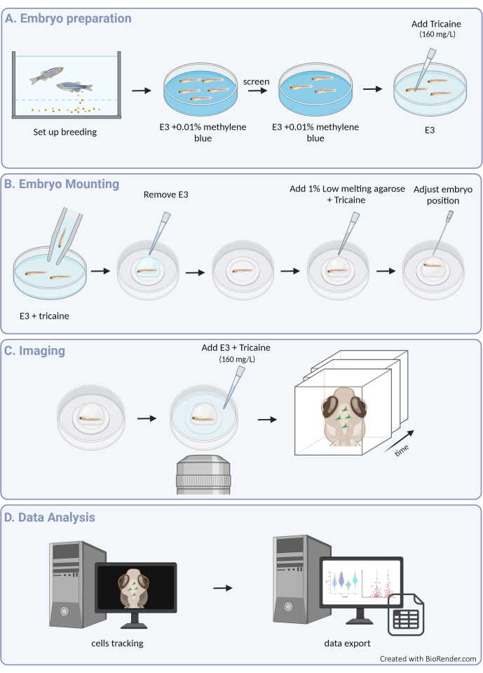
Figure 1: Overview of the experimental procedure. (A) Zebrafish embryo preparation and anesthesia. (B) Sample mounting and positioning. (C) Image acquisition. (D) Image processing and extraction of motility data. Please click here to view a larger version of this figure.
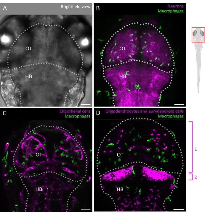
Figure 2: Microglia imaging and interactions with the brain cellular environment in the zebrafish embryo. (A) Brightfield image of a 3 dpf embryonic zebrafish head and brain (dorsal view), labeling the optic tectum and hindbrain. The brain of the embryonic zebrafish can be imaged in its entirety at this stage due to its small size and optical transparency. (B-D) Microglia location and behavior can be visualized by the use of transgenic lines such as (B,C) Tg(mpeg1:eGFP)gl22 and (D) Tg(mpeg1:mcherry)gl23, and focusing on the OT. (B) Neurons and their cell bodies can be identified using the reporter line Tg(XlTubb:DsRed)zf148 and microglia-neuron interactions can be visualized by merging the two channels in lines that co-express both transgenes. Seen is a merge of the green (microglia) and red (neurons, in magenta) signals. (C,D) Reporter lines can also shed light on microglia (in green) interactions with endothelial cells, here labeled in red using the (C) double Tg(kdrl:cres898; actb2:LOXP-STOP-LOXP-Dsredexpress, sd5) transgenics, or with glial cells such as oligodendrocytes and their precursors, using the (D) double Tg(olig2:EGFP; mpeg1:mcherry ) transgenic line. 1: oligodendrocytes and progenitor cells in the OT, 2: eurydendroid neurons in the cerebellum. Scale bars = 50 µm. Abbreviations: dpf = days post fertilization; OT = optic tectum; HB = hindbrain. Please click here to view a larger version of this figure.
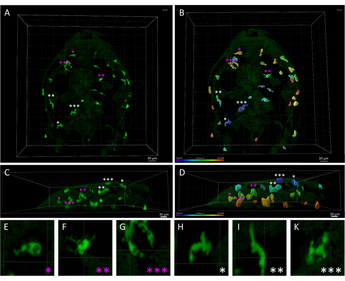
Figure 3: Morphological and spatial disparity between skin macrophages and microglia in a 3 dpf zebrafish embryo. (A,B) Dorsal view of a Tg(mpeg1:eGFP)gl22 zebrafish embryo at 3 dpf, depicting (A) parenchymal microglia cells (marked by magenta asterisks) versus skin macrophages (white asterisks), identified based on their relative position along the z-axis, as seen in (B), which shows the monochrome 3D image rendered with z-axis color-coding. (C,D) Lateral view of (C) A and (D) B, showcasing the z-depths of mpeg1:eGFP+ cells within the embryo's head and highlighting the superficial localization of skin macrophages as compared to microglial cells. (E-K) High magnification of each cell indicated by an asterisk in A and B, providing a detailed visualization of the distinct morphologies between (E-G) amoeboid microglia and (H-K) elongated skin macrophages. Scale bars= 30 µm. Abbreviation: dpf = days post fertilization. Please click here to view a larger version of this figure.
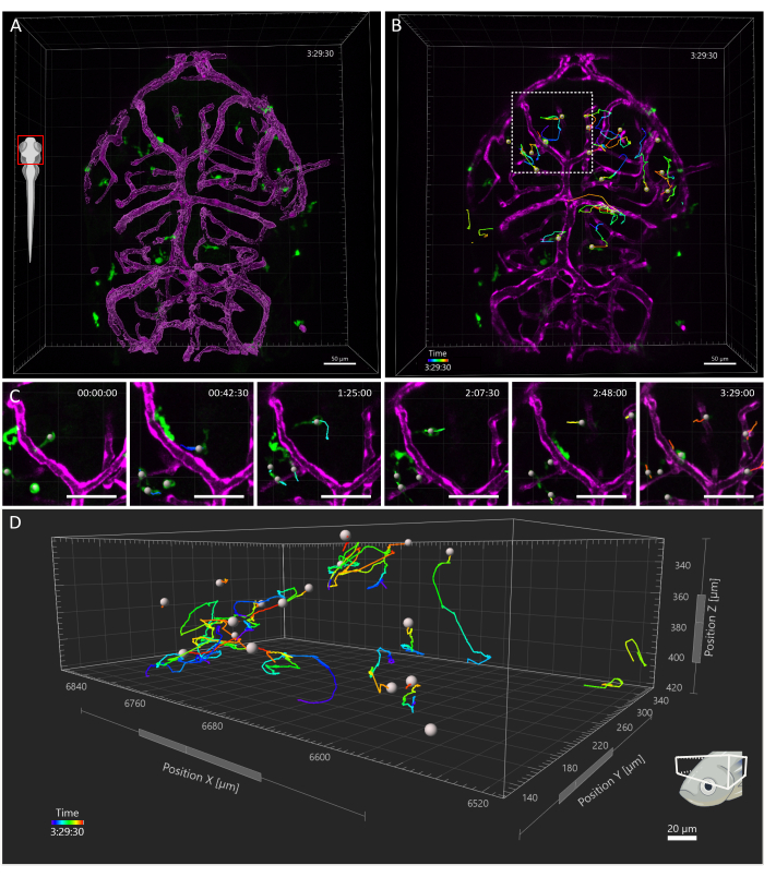
Figure 4: Tracking of microglial cells in a 3 dpf zebrafish embryo. (A) Head dorsal view of a 3 dpf Tg(kdrl:cres898; actb2:LOXP-STOP-LOXP-Dsredexpress,sd5;mpeg1:eGFPgl22) triple transgenic embryo, showing microglia (green) and the surface rendering of the vessels (magenta). (B-D) Representative tracking of microglia movements over a (B) 3.5 h time window. Individual cells are seen to follow complex trajectories. (C) Detail from timelapse, showing a magnified view of the region in B enclosed by the dashed square. Six frames from the movie (45 min apart) are presented, documenting microglia establishing transient contacts with endothelial cells in their microenvironment. (D) Trajectories of individual microglial cell migration within the optic tectum, with the X, Y, and Z axes representing spatial dimensions. Scale bar = 50 µm. Abbreviation: dpf = days post fertilization. Please click here to view a larger version of this figure.
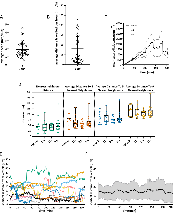
Figure 5: Visualization of the data obtained. Example of the spatial data that can be obtained using the described protocol. The graphs depict (A) the average speed of microglial cells, (B) the average distance they cover in 1 h, (C) their mean square displacement, and (D) their distribution in space at different times. The vessel's surface rendering also allowed for the measurement of the shortest distance between microglia and endothelial cells at any given time, both at (E) single-cell level and as a (F) global average. For A,B, and D, each dot represents an individual cell. N = one embryo. Please click here to view a larger version of this figure.
Supplemental File 1: Please click here to download this File.
Discussion
The current protocol enables in vivo imaging of microglia dynamics in a vertebrate embryo and visualization of the acquired motility data. Microglia colonization of the developing brain occurs very early during embryogenesis and precedes critical events such as peaks of neurogenesis, astrogliogenesis, oligodendrogenesis, and many other cellular processes17. It is therefore not surprising that microglia play important functions in shaping specific aspects of brain development18, for example, through the regulation of neuronal differentiation, migration, and survival19,20,21, as well as synaptic pruning5 and myelination22,23,24.
The contribution of dysfunctional microglia to the pathogenesis and/or progression of neurodevelopmental disorders is also increasingly being recognized25. Indeed, the early presence of microglia in the forming brain exposes these cells to distinct physiological states26 and environmental changes. This can have a significant impact given that microglia are long-lived cells in both rodents and humans, being maintained during the lifespan through self-renewal of local progenitors27,28,29. We believe this protocol could serve as a powerful tool to better characterize the behavior of microglia in these distinct physiological states, as they develop, mature, and establish their network during the successive steps of brain morphogenesis.
Using the setup described here, we have successfully imaged and acquired data on zebrafish larvae as old as 6 dpf. Extending the analysis to later developmental stages will likely succeed but will require adjusting the imaging setup to take into consideration the increased sample size, especially along the z-axis. In attempting this, we suggest focusing on maintaining a low signal-to-noise ratio and fast scanning time, as they are key parameters for a successful analysis.
We suggest a minimum imaging time of 1 h to allow for microglia tracking; the longest imaging window that has been tested with this protocol is 8 h. Moreover, it is important for the tracking analysis to keep the time interval between frames as short as possible, ideally between 30 s and 60 s. This will allow for more accurate and detailed tracking data in downstream analyses. Therefore, especially if detecting more than one fluorophore, it is fundamental to avoid spectral overlap and ensure enough separation between the two fluorophore emission spectra to allow for a simultaneous acquisition, without signal bleedthrough.
Other protocols for high-quality timelapse recording of the zebrafish brain are available30, but this is the first one showing how to successfully track all microglia movement during embryonic development over an extended period. Although the workflow presented here focused on tracking microglia in a physiological context, it can be easily applied to the analysis of microglia in pathology. Indeed, several models of neurodevelopmental disorders, such as autism31, epilepsy32, and schizophrenia33, but also neurodegeneration34 and cancer35, have been established in zebrafish that provide unique opportunities for determining microglial response and behavior in disease conditions.
Notably, this tracking protocol is highly versatile and could also be instrumental for shedding light on the migration patterns of various cell types across diverse anatomical regions of the zebrafish embryo, thus potentially opening up avenues for additional applications, beyond the microglial investigation scope described in this article. Moreover, by harnessing the ability to combine multiple fluorescent transgenic lines, we gain the ability to discern the spatial relationship between microglia and other cell types of the brain microenvironment, with the potential to visualize cellular interactions and cross-talks throughout timelapse recordings, in a non-invasive way. This could be instrumental in unraveling the physiological significance of microglia behavior and contribute to a deeper characterization of these highly motile cells.
Disclosures
The authors declare no conflicts of interest.
Acknowledgements
The authors would like to express their sincere gratitude to Professor Nicolas Bayens for generously providing access to the confocal microscope essential for this study. This work was funded in part by the Funds for Scientific Research (FNRS) under Grant Numbers F451218F and UG03019F, the Alzheimer Research Foundation (SAO-FRA) (to V.W.), A.M. is supported by a Research Fellowship from the FNRS. Figure 1 was created on biorender.com.
Materials
| Name | Company | Catalog Number | Comments |
| 1 L Breeding tanks | Tecniplast | ZB10BTE | |
| 1-phenyl-2-thiourea (PTU) | Sigma-Aldrich | P7629 | Diluted to 0.2 mM in E3 to prevent embryo pigmentation |
| Bottom glass imaging dish | FluoroDish | FD3510-100 | |
| Disposable Graduated transfer pipette | avantor | 16001-188 | |
| Dry block heater | Novolab | Grant QBD4 | To keep low melting agarose at 37 °C |
| Ethyl 3-aminobenzoate methanesulfonate (Tricaine) | Sigma-Aldrich | E10521-50G | |
| Imaris 10.0 | Oxford Instruments | analysis software | |
| Imaris File Converter | Oxford Instruments | https://imaris.oxinst.com/big-data | |
| Laser-scanning confocal microscope | Nikon | Eclipse Ti2-E | |
| Methylene blue | Sigma-Aldrich | M9140-25G | |
| microloader tips | Eppendorf | 5242956003 | |
| NuSieve GTG Agarose | Lonza | 50081 | |
| Petri dishes (90 mm) | avantor | 391-0559 | |
| Pronase | Sigma-Aldrich | 11459643001 | |
| Stainless Steel Forceps Dumont No. 5 | FineScienceTools | 11254-20 | |
| Stereo microscope | Leica | Leica M80 | To mount the embryos |
| teasing needle | avantor | 76549-024 |
References
- Wolf, S. A., Boddeke, H. W., Kettenmann, H. Microglia in physiology and disease. Annu Rev Physiol. 79, 619-643 (2017).
- Nimmerjahn, A., Kirchhoff, F., Helmchen, F. Resting microglial cells are highly dynamic surveillants of brain parenchyma in vivo. Sci Rep. 308 (5726), 1314-1318 (2005).
- Peri, F., Nusslein-Volhard, C. Live imaging of neuronal degradation by microglia reveals a role for v0-ATPase a1 in phagosomal fusion in vivo. Cell. 133 (5), 916-927 (2008).
- Marın-Teva, J. L., et al. Microglia promote the death of developing Purkinje cells. Neuron. 41 (4), 535-547 (2004).
- Paolicelli, R. C., et al. Synaptic pruning by microglia is necessary for normal brain development. Science. 333 (6048), 1456-1458 (2011).
- Hattori, Y. The microglia-blood vessel interactions in the developing brain. Neurosci Res. 187, 58-66 (2023).
- Colonna, M., Butovsky, O. Microglia function in the central nervous system during health and neurodegeneration. Annu Rev Immunol. 35, 441-468 (2017).
- Xu, H. -T., Pan, F., Yang, G., Gan, W. -B. Choice of cranial window type for in vivo imaging affects dendritic spine turnover in the cortex. Nat Neurosci. 10 (5), 549-551 (2007).
- Shin, J., Park, H. -C., Topczewska, J. M., Mawdsley, D. J., Appel, B. Neural cell fate analysis in zebrafish using olig2 BAC transgenics. Methods Cell Sci. 25 (1), 7-14 (2003).
- Chi, N. C., et al. Foxn4 directly regulates tbx2b expression and atrioventricular canal formation. Genes Dev. 22 (6), 734-739 (2008).
- Avdesh, A., et al. Regular care and maintenance of a zebrafish (Danio rerio) laboratory: An introduction. J Vis Exp. (69), e4196(2012).
- Bohnsack, B. L., Gallina, D., Kahana, A. Phenothiourea sensitizes zebrafish cranial neural crest and extraocular muscle development to changes in retinoic acid and IGF signaling. PLoS ONE. 6 (8), e22991(2011).
- Ariga, J., Walker, S. L., Mumm, J. S. Multicolor time-lapse imaging of transgenic zebrafish: visualizing retinal stem cells activated by targeted neuronal cell ablation. J Vis Exp. (43), e2093(2010).
- Ferrero, G., et al. Embryonic microglia derive from primitive macrophages and are replaced by cmyb-dependent definitive microglia in zebrafish. Cell Rep. 24 (1), 130-141 (2018).
- Xu, J., Wang, T., Wu, Y., Jin, W., Wen, Z. Microglia colonization of developing zebrafish midbrain is promoted by apoptotic neuron and lysophosphatidylcholine. Dev Cell. 38 (2), 214-222 (2016).
- Svahn, A. J., et al. Development of ramified microglia from early macrophages in the zebrafish optic tectum. Dev Neurobiol. 73 (1), 60-71 (2013).
- Thion, M. S., Garel, S. Microglial ontogeny, diversity and neurodevelopmental functions. Curr Opin Genet Dev. 65, 186-194 (2020).
- Bohlen, C. J., Friedman, B. A., Dejanovic, B., Sheng, M. Microglia in brain development, homeostasis, and neurodegeneration. Annu Rev Genet. 53, 263-288 (2019).
- Squarzoni, P., et al. Microglia modulate wiring of the embryonic forebrain. Cell Rep. 8 (5), 1271-1279 (2014).
- Schafer, D. P., et al. Microglia sculpt postnatal neural circuits in an activity and complement-dependent. Neuron. 74 (4), 691-705 (2012).
- Parkhurst, C. N., et al. Microglia promote learning-dependent synapse formation through brain-derived neurotrophic factor. Cell. 155 (7), 1596-1609 (2013).
- Green, L. A., O'Dea, M. R., Hoover, C. A., DeSantis, D. F., Smith, C. J. The embryonic zebrafish brain is seeded by a lymphatic-dependent population of mrc1+ microglia precursors. Nat. Neurosci. 25 (7), 849-864 (2022).
- Hughes, A. N., Appel, B. Microglia phagocytose myelin sheaths to modify developmental myelination. Nat Neurosci. 23 (9), 1055-1066 (2020).
- Yu, Q., et al. C1q is essential for myelination in the central nervous system (CNS). iScience. 26 (12), 108518(2023).
- Zengeler, K. E., Lukens, J. R. Innate immunity at the crossroads of healthy brain maturation and neurodevelopmental disorders. Nat Rev Immunol. 21 (7), 454-468 (2021).
- Paolicelli, R. C., et al. Microglia states and nomenclature: A field at its crossroads. Neuron. 110 (21), 3458-3483 (2022).
- Réu, P., et al. The lifespan and turnover of microglia in the human brain. Cell Rep. 20 (4), 779-784 (2017).
- Ajami, B., Bennett, J. L., Krieger, C., Tetzlaff, W., Rossi, F. M. V. Local self-renewal can sustain CNS microglia maintenance and function throughout adult life. Nat Neurosci. 10 (12), 1538-1543 (2007).
- Mildner, A., et al. Microglia in the adult brain arise from Ly-6ChiCCR2+ monocytes only under defined host conditions. Nat Neurosci. 10 (12), 1544-1553 (2007).
- Cook, Z. T., Brockway, N. L., Weissman, T. A. Visualizing the developing brain in living zebrafish using Brainbow and time-lapse confocal imaging. J Vis Exp. (157), e60593(2020).
- Tayanloo-Beik, A., et al. Zebrafish modeling of autism spectrum disorders, current status and future prospective. Front Psychiatry. 13, 911770(2022).
- D'Amora, M., et al. Zebrafish as an innovative tool for epilepsy modeling: State of the art and potential future directions. Int J Mol Sci. 24 (9), 7702(2023).
- Langova, V., Vales, K., Horacek, J. The role of zebrafish and laboratory rodents in schizophrenia research. Front Psychiatry. 11, 549232(2020).
- Chia, K., Klingseisen, A., Sieger, D., Priller, J. Zebrafish as a model organism for neurodegenerative disease. Front Mol Neurosci. 15, 940484(2022).
- Astell, K. R., Sieger, D. Zebrafish in vivo models of cancer and metastasis. Cold Spring Harb Perspect Med. 10 (8), 037077(2020).
Reprints and Permissions
Request permission to reuse the text or figures of this JoVE article
Request PermissionThis article has been published
Video Coming Soon
Copyright © 2025 MyJoVE Corporation. All rights reserved