Method Article
Prion Safety Laboratory Swipe Test
In This Article
Summary
A method to assess commonly used areas in laboratory settings for prion contamination and effective decontamination is lacking. The protocol described here provides key fundamentals for implementing a laboratory prion safety swipe test that can easily be modified to meet the individual needs of specific laboratories.
Abstract
Transmission of iatrogenic prion disease has occurred from contaminated neurosurgical tools, transplant materials, and occupational exposure to prion-contaminated laboratory tools. Prions cause disease by the templated misfolding of the normal cellular form of the prion protein, PrPC, into the misfolded and pathogenic form PrPSc and are invariably fatal. Reducing iatrogenic and occupational prion transmission is challenging. First, prions can bind to and persist on surfaces for long periods of time. Second, prions are highly resistant to inactivation. Given this, surfaces can retain infectivity for long periods of time following ineffective decontamination. Not only can this pose a potential occupational risk for prion laboratory workers, but it could potentially cross-contaminate laboratory experiments utilizing sensitive prion amplification techniques. The protocol described here for a prion safety laboratory swipe test includes steps for the identification and documentation of high-traffic laboratory areas, recommended swabbing controls to ensure the validity of results, steps to identify proper responses to positive surface swabbing sites, representative results from prion swipe testing, as well as potential artifactual results. Overall, the prion safety laboratory swipe test can be implemented as part of a broader prion safety program to assess decontamination of surfaces, monitor common spaces for prion contamination, and implement the documentation of prion decontamination status.
Introduction
Prion diseases are invariably fatal neurodegenerative diseases with no known treatment or cure. Prion diseases are caused by PrPSc, the misfolded and pathogenic form of the normal cellular form of the prion protein1,2,3,4,5, PrPC. Prion diseases are known to affect humans and several other animal species. One human prion disease, Creutzfeldt-Jakob Disease, CJD, has three known etiologies: sporadic, inherited, and acquired. Acquired CJD can occur as a result of accidental transmission (iatrogenic and occupational) and is thought to be the cause of Kuru in the Fore people of Papua New Guinea6.
Prion transmission has been associated with prion-contaminated medical devices and transplant materials7,8,9,10,11,12,13,14,15,16,17. Iatrogenic transmission of CJD can occur via blood, tissue, or from prion-contaminated surfaces18,19,20. For example, iatrogenic CJD can develop in patients following an electroencephalogram with electrodes previously used on an individual in the preclinical stage of CJD who then later succumbed to CJD21. More recent laboratory-based occupational transmission has also occurred where a laboratory worker contracted prion disease via a skin puncture with forceps used to handle brain slices from an animal infected with sheep-adapted BSE22,23. Such transmission scenarios could occur within clinical, laboratory, and diagnostic laboratory settings where prion samples are handled.
Prions resist common disinfection techniques and can persist and remain infectious on surfaces for extended periods of time24,25,26,27,28,29. Common disinfection techniques such as the use of ethanol, phenolic cleaners, hydrogen peroxide, various forms of radiation, and formaldehyde are inadequate for the inactivation of prions, allowing surfaces to retain infectivity30,31,32,33,34,35,36,37. These characteristics contribute to the transmission of prions during iatrogenic and occupational exposure.
Methods for the detection of environmental prions have only recently been developed. An environmental swabbing method coupled with real-time quaking-induced conversion (RT-QuIC) can assess residual prion infectivity from environmental surfaces as well as common laboratory surfaces following ineffective disinfection38,39,40,41,42. Here, we describe how this technique can be incorporated into a broader prion safety program. Overall, this method can allow for the monitoring of laboratory-dependent disinfection protocols, the investigation and proper documentation of contamination status, which can help ensure the validity of experiments by minimizing cross-contamination, the assessment of shared use spaces for prion contamination and allows for directional retraining of personnel based on commonly contaminated areas.
Protocol
All procedures involving animals were approved and in compliance with the Guide for the Care and Use of Laboratory Animals by the Creighton University Institutional Animal Care and Use Committee.
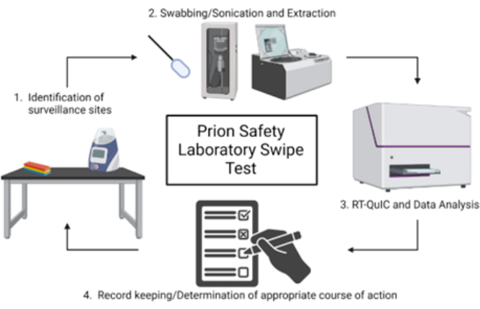
NOTE: A schematic overview of the prion safety laboratory swipe test is shown in Figure 1.
1. Selection of swabbing sites and preparation for surface swabbing
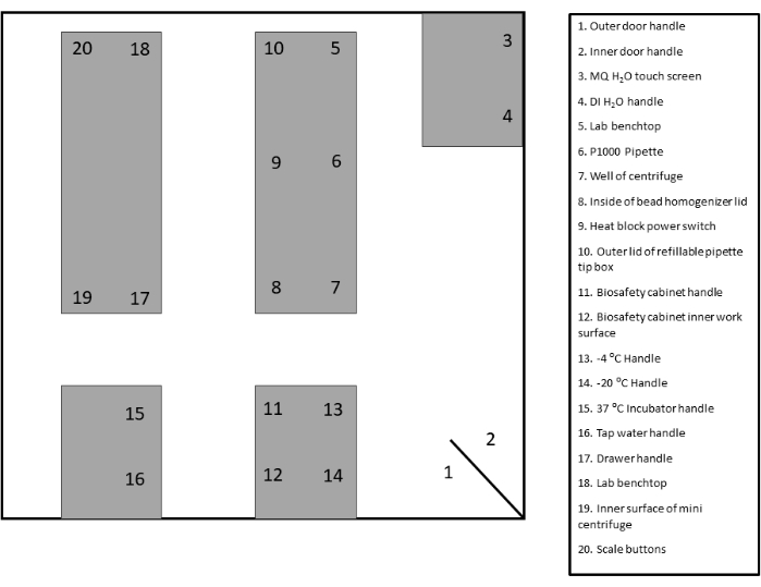
- Identify and label appropriate surveillance areas for swabbing. Refer to the template provided for documentation and reference (Figure 2 and Supplementary Figure 1).
- Prepare two 1.5 mL microcentrifuge tubes for each of the identified areas. Add 250 µL of Dulbecco's phosphate-buffered saline to the first set of microcentrifuge tubes for use in step 3.2. Let the second set of microcentrifuge tubes remain empty for use in step 4.3.
- Retrieve foam-tipped swabs from storage in an area free of prion contamination.
- Prepare a squeeze bottle with Milli-Q water (MQ H2O) for use in step 2.1.
2. Positive and negative control swab preparation
- Prepare dilutions for positive and negative swab controls using relevant control types. Example: For positive control, prepare a 1% dilution of prion-infected brain homogenate in Dulbecco's phosphate-buffered saline (DPBS). For negative control, prepare a 0.1% dilution of uninfected (UN) brain homogenate (BH) from the same species as the positive control in DPBS.
- Using clean gloves, retrieve the appropriate number of swabs from the clean packaging and place the handle side down into a tube rack, taking care to space out foam swabs so that the tips do not contact other swabs or surfaces. For each control sample, prepare three swabs. Change gloves between each sample.
- Holding a clean foam swab by the handle, apply 50 µL of respective positive and negative control samples to foam swab tips. Make sure to apply to both sides of the swab tip to ensure complete absorption.
NOTE: Always change gloves prior to removing a clean swab from the tube rack. - Using scissors, cut off the excess handle of the swab (approximately ½ of the length) and place the swab into the preloaded microcentrifuge tube with the foam tip portion pointed down. Ensure that the foam tip is submerged in the DPBS and that the handle is cut adequately to close the lid of the microcentrifuge tube completely.
- Continue applying control samples after changing gloves until all samples have been applied.
3. Surface swabbing
- Holding a clean foam tip swab by the handle, prewet the foam tip with MQ H2O and shake off the excess. Place the moistened foam tip of the swab onto the area chosen for surveillance and swab the area back and forth approximately ten times while simultaneously rotating the tip of the swab on the surface.
- Using scissors, cut off the excess handle of the swab (approximately ½ of the length) and place the swab into the preloaded microcentrifuge tube with the foam tip portion pointed down. Ensure that the foam tip is submerged in the DPBS and that the handle is cut adequately to close the lid of the microcentrifuge tube completely.
- Discard gloves and place new gloves on before each respective swabbing site to minimize the probability of cross contamination.
- Repeat the procedure until all areas chosen for surveillance have been swabbed.
4. Swab extraction and vacuum concentration
NOTE: Turn on the vacuum concentrator 30 min prior to use to allow the instrument to warm up.
- Place microcentrifuge tubes into a circular tube rack. Place the microfuge tube rack into the cup horn sonicator water bath. Ensure the foam swabs in DPBS within the microcentrifuge tubes are below the surface of the water in the cup horn (the handle portion does not have to be submerged). Apply the following settings: 15 s total run time (5 s on, 5 s off) at ~75-85 watts.
- Following sonication, centrifuge the tubes for approximately 15 s to collect DPBS in the bottom of the tube prior to transfer.
- Using a P1000 pipette set to 250 µL, carefully collect all liquid (swab extract) from the bottom of the microcentrifuge tube and transfer into the corresponding microcentrifuge tube from the second, empty prelabeled set. Use the pipette tip to squeeze any excess liquid from the foam tip. Discard empty tubes containing swabs.
- Set the following settings on the vacuum concentrator: Temperature: 45 °C, Heat time: 15 min, Run time: 2 h, Vacuum: 5.1.
- Place the microcentrifuge tubes containing swab extracts into the vacuum concentrator, ensuring the tubes are balanced and all tube caps are open.
- Upon cycle completion, ensure that samples are completely concentrated (only the pellet remains). Store the pellets at -80 °C until utilized for RT-QuIC.
NOTE: In some instances, additional vacuum concentration time may be required to ensure complete concentration. Remove all concentrated samples, leaving only the tubes that still contain liquid. Rebalance the remaining tubes within the concentrator and run at additional 1-h increments until samples are completely concentrated.
5. Preparation of swabbing controls for use in RT-QuIC
NOTE: The RT-QuIC controls should be performed prior to the assay of environmental swab extracts to ensure that contamination has not been introduced during the swabbing, extraction, or concentration procedures. For example layouts, see Figure 3 and Figure 4.
- Prepare an appropriate negative RT-QuIC plate control (e.g., uninfected brain homogenate [UN BH]) by diluting to the appropriate concentration in tissue dilution solution (N2-0.1%SDS/PBS). Prepare a positive plate control dilution by diluting prion-infected brain homogenate to a dilution of 10-3 in tissue dilution solution.
- Remove previously stored control swab extract pellets from -80 °C and resuspend each with 50 µL of MQ H2O by pipetting up and down approximately 10 times, followed by vortexing briefly. Allow samples to sit at room temperature (RT) while performing the remaining steps.
- Load 2 µL of specified plate controls, including negative plate controls (tissue dilution solution alone and uninfected brain homogenate) and a positive plate control (infected brain homogenate).
- Load 2 µL of each positive and negative swab extract technical replicate into a minimum of four replicate RT-QuIC plate wells.
NOTE: If negative swabbing controls exhibit RT-QuIC seeding above a laboratory's standard for a positive determination, this would indicate that contamination had been introduced during the swabbing, extraction, and concentration process and that the swabbing experiment should be performed again
6. Preparation of samples for use in RT-QuIC
- Prepare an appropriate negative plate control (e.g., uninfected brain homogenate, uninfected lymph node, etc.) by diluting to the appropriate concentration in tissue dilution solution (N2-0.1%SDS/PBS). Prepare positive plate controls by diluting infected brain homogenate to a dilution of 10-3 in tissue dilution solution.
- Remove previously stored swab extract pellets from -80 °C and resuspend with 20-50 µL of tissue dilution solution (depending on the suspected amount of contamination) by pipetting up and down approximately ten times, followed by vortexing briefly. Allow samples to sit at RT while performing the remaining steps.
- Load 2 µL of specified plate controls, including negative plate controls (tissue dilution solution alone and uninfected brain homogenate) and a positive plate control (infected brain homogenate) into the RT-QuIC plate wells.
- Load 2 µL of each swab extract into four replicate wells.
7. RT-QuIC analysis and results
- Perform RT-QuIC according to individual laboratory protocols (optional protocols38,40).
- Define the parameters required for determining the positivity of a sample (see representative results, Figure 5).
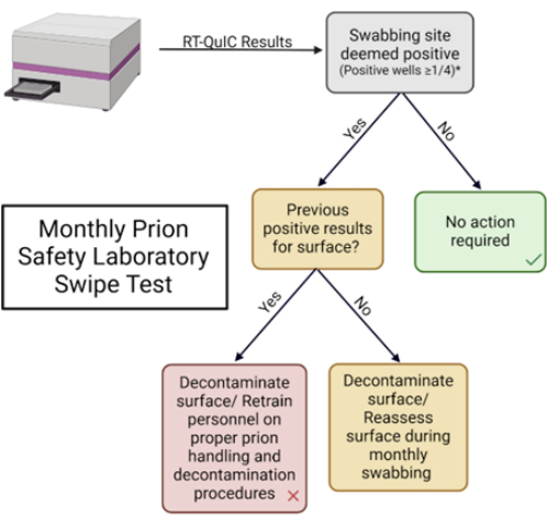
NOTE: These parameters are defined by each lab. - Record results in the provided table and take appropriate action based on laboratory best practices (Table 1 and Supplementary Figure 2).
Results
Written description of positive and negative results (including positive/negative plate and swab controls)
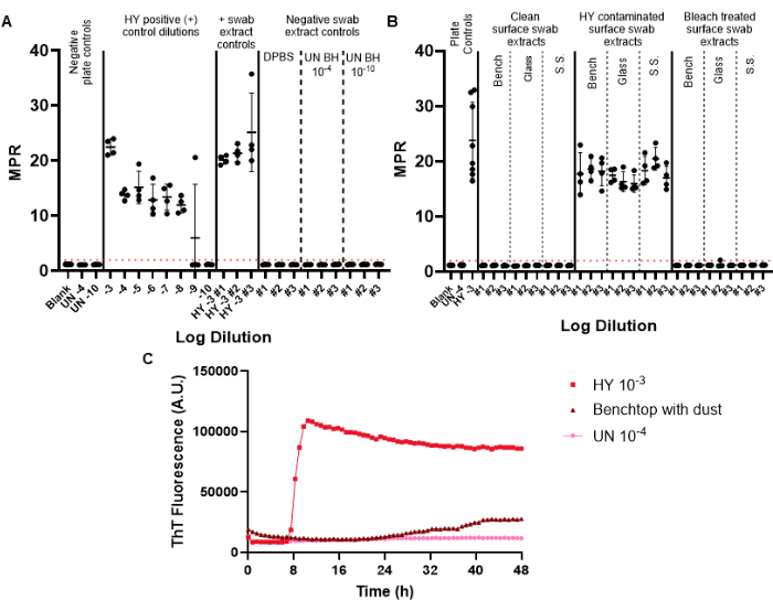
Negative control swabs are included in the surveillance swabbing to monitor for potential prion contamination that could be introduced during the swabbing, extraction, and concentration process. The first RT-QuIC plate performed for a given monthly surveillance should include the positive and negative swab controls. Successful negative controls fail to cross the positive fluorescence threshold (Figure 6A). This result would indicate that contamination had not been introduced during the experimental procedures. Successful positive control swab extracts would exhibit positive seeding in all replicate wells for a given sample (positive control swabs were contaminated with the hamster-adapted transmissible mink encephalopathy strain Hyper (HY TME) brain homogenate. The inclusion of a positive control dilution series allows for the determination of the sensitivity of prion detection for a given experiment (Figure 6A).
When examining surface swab extract samples, surfaces that fail to show seeding above the predetermined positive fluorescence threshold can be considered to be prion negative (Figure 6B). Conversely, prion-contaminated surface swab extracts will show seeding capabilities above the positive fluorescence threshold, although the maxpoint ratio (MPR) and time to fluorescence can vary compared to the included positive plate control (Figure 6B). The ability of the method to assess adequate disinfection is highlighted by the bleach-treated prion contaminated surfaces, which now fail to seed RT-QuIC (Figure 6B).
Importantly, while our laboratory defines a positive sample as a sample that passes the set positive fluorescence threshold in at least half of the replicate wells, it is necessary for each laboratory to set its own standards. The rate of amyloid formation (RAF) and time to fluorescence can also be used to help establish laboratory-specific thresholds for positivity.
We have observed surface artifact results that pass the positive fluorescence threshold but with altered kinetic curves and after an abnormally long time to fluorescence compared to positive control samples (Figure 6C). These results should be cautiously interpreted as they may be caused by the presence of dust or residual chemicals present on a surface. These findings highlight the necessity of general laboratory cleanliness, as well as criteria for differentiating between true positive and false positive seeding.
Disposal of solid and liquid biohazard waste should be done following a given institution's current biohazard disposal guidelines. Common prion disinfection techniques include treatment with sodium hydroxide, sodium hypochlorite (bleach), or autoclaving at 134 °C for 18 min43,44,45,46.

Figure 1: Schematic overview of prion safety laboratory swipe test. Please click here to view a larger version of this figure.

Figure 2: Sample swabbing site layout. Please click here to view a larger version of this figure.

Figure 3: Sample experiment design for swab controls included prion safety laboratory swipe test. (A) Sample layout for negative and positive swab extract controls. (B) Negative and positive swab extract controls should be resuspended with 50 µL of H2O. A 10-fold dilution of the resuspended swab extract should be generated by diluting it into a tissue dilution solution. Please click here to view a larger version of this figure.

Figure 4: Sample experiment design for surface swab extracts from prion safety laboratory swipe test. (A) Sample layout for surface swab extracts. (B) Surface swab extract samples should be resuspended with 20 µL of tissue dilution solution. Please click here to view a larger version of this figure.

Figure 5: Monthly prion safety laboratory swipe test. Flowchart used for interpreting results and determining appropriate response. Please click here to view a larger version of this figure.

Figure 6: Representative surface swabbing experiment. (A) Surface swab control plate containing negative swab extract controls in triplicate (DPBS, uninfected hamster brain homogenate 10-4 and 10-10) and positive swab extract controls in triplicate (HY brain homogenate 10-3). (B) Representative swab extracts for benchtop, glass, and stainless steel (S.S.) surfaces that were swabbed prior to contamination, following contamination with HY 10-3, and following bleach treatment of contaminated surfaces. (C) Fluorescence tracing comparison of uninfected brain homogenate 10-4, HY brain homogenate 10-3, and the swab extract from the benchtop coated with a fine film of dust. Negative plate controls for panels A and B include a blank (tissue dilution solution) and uninfected hamster brain homogenate 10-4. Panel B includes the addition of uninfected brain homogenate 10-4 and the positive plate control of HY brain homogenate 10-3. A positive fluorescence threshold (illustrated by a red dashed line) was determined to be at 2. The maxpoint ratio (MPR) reported is the maximum fluorescence divided by the initial fluorescence reading obtained by the plate reader. Each point represents one technical well replicate for a given sample type. The mean and standard deviation are presented. Please click here to view a larger version of this figure.
Table 1: Sample swabbing site monthly documentation form. Please click here to download this Table.
Supplementary Figure 1: Swabbing site layout template. Please click here to download this File.
Supplementary Figure 2: Swabbing site monthly documentation form. Please click here to download this File.
Discussion
The described prion safety swabbing method can be used to enhance existing prion safety measures. This method can monitor prion laboratory spaces and equipment, as well as shared laboratory spaces, for potential prion contamination. Importantly, this method can be adapted to test laboratory-specific disinfection techniques to verify decontamination of prion-contaminated surfaces. As various prion strains display different sensitivities to disinfection techniques, this method can confirm that these techniques are effective for current laboratory experiments, such as treatment with sodium hydroxide or sodium hypochlorite (bleach)43,44.
Key steps for this methodology include the identification of appropriate swabbing sites that will provide a sample of both high-traffic areas as well as areas to monitor that may be involved with downstream cross-contamination of experiments. Additionally, laboratories should utilize positive and negative controls that closely mirror those commonly used in a given lab or clinic. For example, if working with rodent prions, the positive and negative controls should match that species. Finally, surveillance data should be kept up to date and organized so that trends in positivity can easily be identified, thus allowing for mitigation and retraining of laboratory staff in the event of consistent positive results for a given area.
One limitation of this protocol is the potential generation of erroneous RT-QuIC results. Given the sensitive nature of RT-QuIC, the presence of residual detergents, salt, and other substances on laboratory surfaces can impact the outcome of the reaction39. Overall, given this observation, it is advantageous that surfaces remain free of substances that can interfere with RT-QuIC, such as residual salt, detergents, and dust. As improvements to the RT-QuIC methodology become available, we anticipate that many of the current limitations of RT-QuIC will be resolved. We, therefore, encourage the end user to keep abreast of the literature to take advantage of the latest improvements in RT-QuIC.
A key benefit of this method for laboratories researching prions is the ability to minimize contamination that may impact the results of sensitive amplification assays. Necropsy tools are commonly disinfected prior to being reused for future necropsies. The described swabbing method provides a method to assess necropsy tools for residual prion infectivity that may affect downstream results. This can provide an additional level of rigor to experiments to exclude the possibility that prion detection in tissues was not due to contamination from the necropsy tools.
Disclosures
J.C.B. and Q.Y. are inventors on a patent application pertaining to prion surface swabbing technology.
Acknowledgements
The work was supported by a grant from the Creutzfeldt Jacob Disease Foundation. The funders had no role in study design, data collection, and interpretation, or the decision to submit the work for publication.
Materials
| Name | Company | Catalog Number | Comments |
| Fisherbrand PurSwab Foam Swabs | Fisher brand | Catalog #14-960-3E | |
| Milli-Q IQ 7005 Ultrapure Water System | MilliporeSigma | Q7005T0C | |
| Mini-centrifuge, 6000 rpm | Southern Labware | MLX-306 | |
| Omega plate reader | BMG Labtech | FLUOstar Omega plate reader | |
| Q700 sonicator | QSonica | Q700-110 | |
| Recombinant hamster prion protein | MNPRO | MNPROtein-Hamster | syrian hamster, amino acids 90-231 |
| Savant speedvac | Thermo Scientific | SPD1030-230 | |
| Silica nanospheres (50 nm) | nano Composix | SISN50-25M |
References
- Prusiner, S. B. Novel proteinaceous infectious particles cause scrapie. Science. 216 (4542), 136-144 (1982).
- Bolton, D. C., McKinley, M. P., Prusiner, S. B. Identification of a protein that purifies with the scrapie prion. Science. 218 (4579), 1309-1311 (1982).
- Oesch, B., et al. A cellular gene encodes scrapie PrP 27-30 protein. Cell. 40 (4), 735-746 (1985).
- Caughey, B., Raymond, G. The scrapie-associated form of PrP is made from a cell surface precursor that is both protease- and phospholipase-sensitive. J Biol Chem. 266 (27), 18217-18223 (1991).
- Deleault, N. R., Harris, B. T., Rees, J. R., Supattapone, S. Formation of native prions from minimal components in vitro. Proc Natl Acad Sci U S A. 104 (23), 9741-9746 (2007).
- Gajdusek, D. C., Zigas, V. Degenerative disease of the central nervous system in New Guinea: the endemic occurrence of "kuru" in the native population. N Engl J Med. 257, 974-978 (1957).
- Centers for Disease Control. Fatal degenerative neurologic disease in patients who received pituitary-derived human growth hormone. MMWR Morb Mortal Wkly Rep. 34, 359-361 (1985).
- Duffy, P., Wolf, J., Collins, G., DeVoe, A. G., Streeten, B., Cowen, D. Letter: possible person-to-person transmission of Creutzfeldt-Jakob disease. N Engl J Med. 290 (12), 692-693 (1974).
- Brown, P., et al. Iatrogenic Creutzfeldt-Jakob disease at the millennium. Neurology. 55 (8), 1075-1081 (2000).
- Huillard d'Aignaux, J., et al. Incubation period of Creutzfeldt-Jakob disease in human growth hormone recipients in France. Neurology. 53 (6), 1197-1201 (1999).
- Marzewski, D. J., Towfighi, J., Harrington, M. G., Merril, C. R., Brown, P. Creutzfeldt-Jakob disease following pituitary-derived human growth hormone therapy: a new American case. Neurology. 38, 1131-1133 (1988).
- Hannah, E. L., et al. Creutzfeldt-Jakob disease after receipt of a previously unimplicated brand of dura mater graft. Neurology. 56 (8), 1080-1083 (2001).
- Shimizu, S., et al. Creutzfeldt-Jakob disease with florid-type plaques after cadaveric dura mater grafting. Arch Neurol. 56 (3), 357-362 (1999).
- Antoine, J. C., et al. Creutzfeldt-Jakob disease after extracranial dura mater embolization for a nasopharyngeal angiofibroma. Neurology. 48 (5), 1451-1453 (1997).
- Esmonde, T., Lueck, C. J., Symon, L., Duchen, L. W., Will, R. G. Creutzfeldt-Jakob disease and lyophilised dura mater grafts: report of two cases. J Neurol Neurosurg Psychiatry. 56, 999-1000 (1993).
- Willison, H. J., Gale, A. N., McLaughlin, J. E. Creutzfeldt-Jakob disease following cadaveric dura mater graft. J Neurol Neurosurg Psychiatry. 54, 940(1991).
- Diringer, H., Braig, H. R. Infectivity of unconventional viruses in dura mater. Lancet. 334, 439-440 (1989).
- Peden, A. H., Head, M. W., Ritchie, D. L., Bell, J. E., Ironside, J. W. Preclinical vCJD after blood transfusion in a PRNP codon 129 heterozygous patient. Lancet. 364 (9433), 527-529 (2004).
- Will, R. G., Matthews, W. B. Evidence for case-to-case transmission of Creutzfeldt-Jakob disease. J Neurol Neurosurg Psychiatry. 45, 235-238 (1982).
- el Hachimi, K. H., Chaunu, M. P., Cervenakova, L., Brown, P., Foncin, J. F. Putative neurosurgical transmission of Creutzfeldt-Jakob disease with analysis of donor and recipient: agent strains. C R Acad Sci III. 320 (4), 319-328 (1997).
- Alper, T., et al. Does the agent of scrapie replicate without nucleic acid. Nature. 214, 764-766 (1967).
- Casassus, B. France halts prion research amid safety concerns. Science. 373 (6554), 475-476 (2021).
- Brandel, J. P., et al. Variant Creutzfeldt-Jakob disease diagnosed 7.5 years after occupational exposure. N Engl J Med. 383 (1), 83-85 (2020).
- Flechsig, E., Hegyi, I., Enari, M., Schwarz, P., Collinge, J., Weissmann, C. Transmission of scrapie by steel-surface-bound prions. Mol Med. 7 (10), 679-684 (2001).
- Zobeley, E., Flechsig, E., Cozzio, A., Enari, M., Weissmann, C. Infectivity of scrapie prions bound to a stainless steel surface. Mol Med. 5 (4), 240-243 (1999).
- Brown, P., Gajdusek, D. C. Survival of scrapie virus after 3 years' interment. Lancet. 337 (8736), 269-270 (1991).
- Seidel, B., et al. Scrapie agent (strain 263K) can transmit disease via the oral route after persistence in soil over years. PLoS One. 2 (5), e435(2007).
- Maddison, B. C., et al. Environmental sources of scrapie prions. J Virol. 84 (21), 11560-11562 (2010).
- Pritzkow, S., Morales, R., Lyon, A., Concha-Marambio, L., Urayama, A., Soto, C. Efficient prion disease transmission through common environmental materials. J Biol Chem. 293 (9), 3363-3373 (2018).
- Alper, T., Cramp, W., Haig, D., Clarke, M. Does the agent of scrapie replicate without nucleic acid. Nature. 214 (90), 764-766 (1967).
- Bernoulli, C., et al. Danger of accidental person-to-person transmission of Creutzfeldt-Jakob disease by surgery. Lancet. 309 (8009), 478-479 (1977).
- Brown, P., Rohwer, R. G., Green, E. M., Gajdusek, D. C. Effect of chemicals, heat, and histopathologic processing on high- infectivity hamster-adapted scrapie virus. J Infect Dis. 145 (5), 683-687 (1982).
- Brown, P., et al. Chemical disinfection of Creutzfeldt-Jakob disease virus. N Engl J Med. 306, 1279-1282 (1982).
- Dickinson, A. G., Taylor, D. M. Resistance of scrapie agent to decontamination. N Engl J Med. 299, 1413-1414 (1978).
- Hunter, G. D., Millson, G. C. Studies on the heat stability and chromatographic behavior of the scrapie agent. J Gen Microbiol. 37, 251-258 (1964).
- Pattison, I. H. Resistance of the scrapie agent to formalin. J Comp Pathol. 75, 159-164 (1965).
- Fraser, H., Farquhar, C., McConnell, I., Davies, D. The scrapie disease process is unaffected by ionising radiation. Prog Clin Biol Res. 317, 653-658 (1989).
- Yuan, Q., et al. Sensitive detection of chronic wasting disease prions recovered from environmentally relevant surfaces. Environ Int. 166, 107347(2022).
- Simmons, S. M., et al. Rapid and sensitive determination of residual prion infectivity from prion-decontaminated surfaces. mSphere. 9, e00504-e00524 (2024).
- Orru, C. D., et al. Sensitive detection of pathological seeds of alpha-synuclein, tau and prion protein on solid surfaces. PLoS Pathog. 20 (4), e1012175(2024).
- Wilham, J. M., et al. Rapid end-point quantitation of prion seeding activity with sensitivity comparable to bioassays. PLoS Pathog. 6 (12), e1001217(2010).
- Srivastava, A., et al. Enhanced quantitation of pathological alpha-synuclein in patient biospecimens by RT-QuIC seed amplification assays. PLoS Pathog. 20 (9), e1012554(2024).
- Hughson, A. G., et al. Inactivation of prions and amyloid seeds with hypochlorous acid. PLoS Pathog. 12 (9), e1005914(2016).
- Williams, K., Hughson, A. G., Chesebro, B., Race, B. Inactivation of chronic wasting disease prions using sodium hypochlorite. PLoS One. 14 (10), e0223659(2019).
- Taylor, D. M. Autoclaving standards for Creutzfeldt-Jakob disease agent [letter]. Ann Neurol. 22 (4), 557-558 (1987).
- Brown, P., Rohwer, R. G., Gajdusek, D. C. Newer data on the inactivation of scrapie virus or Creutzfeldt-Jakob disease virus in brain tissue. J Infect Dis. 153 (6), 1145-1148 (1986).
Reprints and Permissions
Request permission to reuse the text or figures of this JoVE article
Request PermissionThis article has been published
Video Coming Soon
Copyright © 2025 MyJoVE Corporation. All rights reserved