
JoVE 비디오를 활용하시려면 도서관을 통한 기관 구독이 필요합니다. 전체 비디오를 보시려면 로그인하거나 무료 트라이얼을 시작하세요.
Method Article
세포외 플럭스 분석을 이용한 단일 3D 미세조직 구상체의 미토콘드리아 에너지 대사 탐구
Erratum Notice
요약
이 프로토콜은 사용자가 Seahorse 세포외 플럭스 분석을 사용하여 3D 암 세포주 유래 구상체에서 미토콘드리아 에너지 대사를 조사하는 데 도움이됩니다.
초록
구상체라고 불리는 3차원 (3D) 세포 응집체는 최근 몇 년 동안 시험관 내 세포 배양의 최전선이되었습니다. 세포를 2차원, 단세포 단층(2D 배양)으로 배양하는 것과는 달리, 스페로이드 세포 배양은 세포외 매트릭스 단백질의 발현, 세포 신호전달, 유전자 발현, 단백질 생산, 분화 및 증식을 포함하여 생체 내에 존재하는 생리적 세포 구조 및 특성을 촉진, 조절 및 지원한다. 3D 배양의 중요성은 종양학, 당뇨병, 줄기 세포 생물학 및 조직 공학을 포함한 많은 연구 분야에서 인정 받고 있습니다. 지난 십 년 동안 구상체를 생산하고 신진 대사 기능과 운명을 평가하기 위해 개선 된 방법이 개발되었습니다.
세포외 플럭스(XF) 분석기는 XF24 섬 포획 플레이트 또는 XFe96 스페로이드 마이크로플레이트를 사용하여 구상체와 같은 3D 미세조직에서 미토콘드리아 기능을 탐색하는 데 사용되었습니다. 그러나, XF 기술을 이용한 구상체에서 미토콘드리아 에너지 대사를 조사하는 뚜렷한 프로토콜과 최적화는 상세히 설명되지 않았다. 이 백서에서는 XFe96 XF 분석기가 있는 스페로이드 마이크로플레이트를 사용하여 단일 3D 구상체에서 미토콘드리아 에너지 대사를 조사하기 위한 상세한 프로토콜을 제공합니다. XF 기술은 서로 다른 암 세포주를 사용하여 크기가 다른 것뿐만 아니라 다른 부피, 세포 번호, DNA 함량 및 유형의 3D 구상체에서 세포 호흡을 구별 할 수있는 것으로 입증되었습니다.
올리고마이신, BAM15, 로테논 및 안티마이신 A의 최적의 미토콘드리아 이펙터 화합물 농도는 3D 구상체에서 미토콘드리아 에너지 대사의 특정 파라미터를 조사하는 데 사용됩니다. 이 백서에서는 또한 구상체에서 얻은 데이터를 정상화하는 방법에 대해 논의하고 XF 기술을 사용하여 스페로이드 대사를 탐구 할 때 고려해야 할 많은 고려 사항을 다룹니다. 이 프로토콜은 고급 시험관 내 스페로이드 모델에 대한 연구를 추진하는 데 도움이됩니다.
서문
생물학적 연구에서 시험관 내 모델의 발전은 지난 20 년 동안 빠르게 진행되었습니다. 이러한 모델에는 이제 오르간-온-칩 양식, 오가노이드 및 3D 미세조직 구상체가 포함되며, 이들 모두는 시험관내 및 생체내 연구 사이의 번역을 개선하기 위한 공통적인 초점이 되었다. 고급 시험관 내 모델, 특히 구상체의 사용은 조직 공학, 줄기 세포 연구, 암 및 질병 생물학 1,2,3,4,5,6,7 및 유전 독성 학 8,9,10, 나노 물질 독성학 11을 포함한 안전성 테스트를 포함한 여러 연구 분야에 걸쳐 있습니다
Access restricted. Please log in or start a trial to view this content.
프로토콜

그림 1: 세포 구상체 생성을 위한 그래픽 워크플로우, 세포외 플럭스 분석 및 다운스트림 분석. 네 개의 암 세포주를 단층(A)으로 선택적으로 배양하고, 조직 배양 플라스크로부터 분리하고, 초저 부착 96-웰 마이크로플레이트에 시딩하여 구상체(B)를 형성하였다. A549 폐 암종, HepG2/C3A 간 암종, SK-OV-3 난소 선암종 및 MCF-7 유방 암종 세포를 1 × 10 3-8 × 10³ 세포/웰에서 시딩하고 7일까지 성장시켜 단일 구상체를 형성하고 지속적인 관찰 및 평면 측정을 통해 스페로이드 시딩 밀도 및 배양 시간을 최적화하였다. 일단 형성되면, 단일 구상체를 무혈청 XF 배지로 세척하고, 폴리-D-리신 (C)으로 예비코팅된 스페로이드 분석 마이크로플레이트에 조심스럽게 시딩하였....
Access restricted. Please log in or start a trial to view this content.
결과
잘 형성되고 컴팩트한 구상체를 얻기 위해, 각 세포주는 시딩 밀도 및 배양 기간 동안 개별적으로 최적화되었다(도 3). A549, HepG2/C3A 및 SK-OV-3 세포주는 초기에 느슨한 응집체를 형성하였고, 이는 배양 7일 후까지 명확하게 정의된 둘레를 갖는 둥근 구상체로 진행되지 않았다. 반대로, MCF-7 세포는 3일 이내에 구상체를 형성할 수 있었다. 모든 스페로이드 모델에 대한 배양 기간 ?.......
Access restricted. Please log in or start a trial to view this content.
토론
주요 결과 및 결과
이 논문은 XFe96 XF 분석기와 함께 일련의 암 유래 세포주를 사용하여 단일 3D 구상체의 미토콘드리아 에너지 대사를 조사하는 상세한 프로토콜을 제공한다. 강제 응집을 위한 세포 기피제 기술을 사용하여 A549, HepG2/C3A, MCF7 및 SK-OV-3 세포 구상체의 신속한 배양을 위한 방법이 개발되고 기술된다. 이 프로토콜은 (1) 스페로이드 배양 프로토콜의 최적화 및 기술 제조업?.......
Access restricted. Please log in or start a trial to view this content.
공개
저자는 선언 할 이해 상충이 없습니다.
감사의 말
N.J.C는 Sygnature Discovery Ltd (BB/M01116X/1, 1940003)와 함께 BBSRC MIBTP CASE Award의 지원을 받았습니다.
....Access restricted. Please log in or start a trial to view this content.
자료
| Name | Company | Catalog Number | Comments |
| A549 | ECACC | #86012804 | Lung carcinoma cell line |
| Agilent Seahorse XF RPMI Medium, pH 7.4 | Agilent Technologies Inc. | 103576-100 | XF assay medium with 1 mM HEPES, without phenol red, sodium bicarbonate, glucose, L-glutamine, and sodium pyruvate |
| Agilent Seahorse XFe96 Extracellular Flux Analyzer | Agilent Technologies Inc. | - | Instrument for measuring rates of spheroid oxygen uptake in single spheroids |
| Antimycin A | Merck Life Science | A8674 | Mitochondrial respiratory complex III inhibitor |
| BAM15 | TOCRIS bio-techne | 5737 | Mitochondrial protnophore uncoupler |
| Black-walled microplate | Greiner Bio-One | 655076 | For fluorescence-based assays |
| CELLSTAR cell-repellent surface 96 U well microplates | Greiner Bio-One | 650970 | Microplates for generating spheroids |
| CellTiter-Glo 3D Cell Viability Assay | Promega | G9681 | Assay for the determination of cell viability in 3D microtissue spheroids |
| Cultrex Poly-D-Lysine | R&D Systems a biotechne brand | 3439-100-01 | Molecular cell adhesive for coating XFe96 spheroid microplates to facillitate attachment of spheroids |
| D-(+)-Glucose | Merck Life Sciences | G8270 | Supplement for cell culture growth and XF assay medium |
| Dulbecco’s Modified Eagle Medium (DMEM) | Gibco | 11885084 | Culture medium for HepG2/C3A spheroids |
| EVOS XL Core Imaging System | Thermo Fisher Scientific | AMEX1000 | Phase-contrast imaging microscope |
| EZ-PCR Mycoplasma test kit | Biological Industries | 20-700-20 | Mycoplasma screening in cell cultures |
| FIJI Is Just Image J | Analysis of collated images | ||
| Foetal bovine serum | Merck Life Science | F7524 | Supplement for cell culture medium |
| HepG2/C3A | ATCC | #CRL-10741 | Hepatic carcinoma cell line, a clonal derivative of the parent HepG2 cell line |
| Lactate-Glo | Promega | J5021 | Assay for measurement of lactate within spheorid culture medium |
| L-glutamine (200 mM solution) | Merk Life Sciences | G7513 | Supplement for cell culture growth and XF assay medium |
| M50 Stereo microscope | Leica Microsytems | LEICAM50 | Stereo dissection micrscope; used for spheorid handling |
| MCF-7 | ECACC | #86012803 | Breast adenocarcinoma cell line |
| Oligomycin from Streptomyces diastatochromogenes | Merck Life Science | O4876 | ATP Synthase Inhibitor |
| Penicilin-Streptomycin | Gibco | 15140122 | Antibiotics added to cell culture medium |
| Quant-iT PicoGreen dsDNA Assay Kit | Initrogen | P7589 | Analysis of dsDNA in spehroids |
| Rotenone | Merck Life Science | R8875 | Mitochondrial Respiratory Complex I Inhibitor |
| RPMI 1640 | Gibco | 21875091 | Culture medium for A549, MCF7, and SK-OV-3 spheroids |
| Seahorse Analytics | Agilent Technologies Inc. | Build 421 | https://seahorseanalytics.agilent.com |
| Seahorse XFe96 Spheroid FluxPak | Agilent Technologies Inc. | 102905-100 | Each Seahorse XFe96 Spheroid FluxPak contains: 6 Seahorse XFe96 Spheroid Microplates (102978-100), 6 XFe96 sensor cartridges, and 1 bottle of Seahorse XF Calibrant Solution 500 mL (100840-000) |
| Serological pipette: 5, 10, and 25 mL | Greiner Bio-One | 606107; 607107; 760107 | Consumables for cell culture |
| SK-OV-3 | ECACC | #HTB-77 | Ovarian adenocarcinoma cell line |
| Sodium pyruvate (100 mM solution) | Merck Life Science | S8636 | Supplement for cell culture growth and XF assay medium |
| T75 cm2 cell culture flask | Greiner Bio-One | 658175 | Tissue culture treated flasks for maintaining cell cultures |
| TrypLExpress | Gibco | 12604-021 | Cell dissociation reagent |
| Wave controller software | Agilent Technologies Inc. | - | |
| Wide orifice tip | STARLAB International GmbH | E1011-8400 | Pipette tips with wide opening for spheroid handling |
참고문헌
- Correa de Sampaio, P., et al. A heterogeneous in vitro three dimensional model of tumour-stroma interactions regulating sprouting angiogenesis. PLoS One. 7 (2), 30753(2012).
- Amann, A., et al.
Access restricted. Please log in or start a trial to view this content.
Erratum
Formal Correction: Erratum: Exploring Mitochondrial Energy Metabolism of Single 3D Microtissue Spheroids using Extracellular Flux Analysis
Posted by JoVE Editors on 3/11/2022. Citeable Link.
An erratum was issued for: Exploring Mitochondrial Energy Metabolism of Single 3D Microtissue Spheroids using Extracellular Flux Analysis. The Representative Results section was updated.
Figure 5 was updated from:
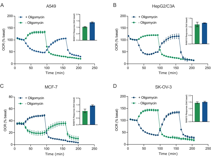
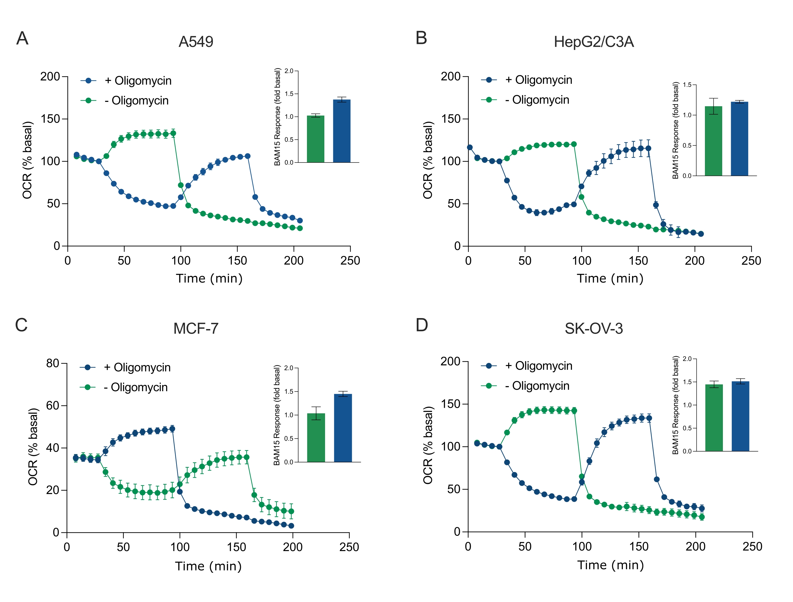
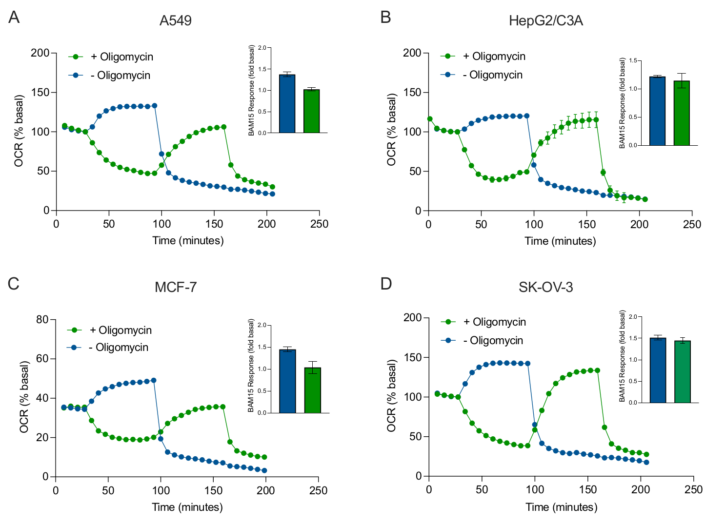
Figure 5: Single or sequential injection of mitochondrial respiratory compounds. Cancer-cell-derived spheroids of MCF-7, HEPG2/C3A, SK-OV-3, and A549 were placed into wells of an XFe96 spheroid microplate in XF RPMI and probed for OCR using the Agilent Seahorse XFe96 analyzer. OCR was measured 5x, after which 2 µg/mL oligomycin (injection Port A: green trace) or 5 µM BAM15 (injection Port A: blue trace or injection port B: green trace) to inhibit the mitochondrial ATP synthase and determine maximal respiratory capacity, respectively. Kinetic OCR data are expressed as % basal (A-D). Maximal respiratory capacity (OCRmax) was calculated as a factor of basal OCR by the equation: OCRmax = OCRBAM15 / OCRbasal. OCRmax was obtained from OCR averages across measurement cycles 8-10 post BAM15 injection with (green bars) and without (blue bars) oligomycin. Data are averages ± SEM from 3-8 individual well replicates across the spheroid assay microplate. Abbreviations: OCR = oxygen consumption rate. Please click here to view a larger version of this figure.
to:

Figure 5: Single or sequential injection of mitochondrial respiratory compounds. Cancer-cell-derived spheroids of MCF-7, HEPG2/C3A, SK-OV-3, and A549 were placed into wells of an XFe96 spheroid microplate in XF RPMI and probed for OCR using the Agilent Seahorse XFe96 analyzer. OCR was measured 5x, after which 2 µg/mL oligomycin (injection Port A: green trace) or 5 µM BAM15 (injection Port A: blue trace or injection port B: green trace) to inhibit the mitochondrial ATP synthase and determine maximal respiratory capacity, respectively. Kinetic OCR data are expressed as % basal (A-D). Maximal respiratory capacity (OCRmax) was calculated as a factor of basal OCR by the equation: OCRmax = OCRBAM15 / OCRbasal. OCRmax was obtained from OCR averages across measurement cycles 8-10 post BAM15 injection with (green bars) and without (blue bars) oligomycin. Data are averages ± SEM from 3-8 individual well replicates across the spheroid assay microplate. Abbreviations: OCR = oxygen consumption rate. Please click here to view a larger version of this figure.
재인쇄 및 허가
JoVE'article의 텍스트 или 그림을 다시 사용하시려면 허가 살펴보기
허가 살펴보기더 많은 기사 탐색
This article has been published
Video Coming Soon
Copyright © 2025 MyJoVE Corporation. 판권 소유