Method Article
Vitrification of Ovarian Cortex Tissue to Achieve a Glassy State of Aggregation
In This Article
Summary
A protocol for the vitrification of ovarian tissue, as an alternative cryopreservation method to the widely used slow freezing protocol, is presented.
Abstract
Ovarian tissue cryopreservation (OTC) is an important option for fertility preservation. For patients whose gonadotoxic treatments cannot be postponed or for pre-pubertal girls, it is often the only option for fertility protection. Cryopreservation can be performed either by vitrification or by slow freezing. Slow freezing is currently the standard approach. An increasing number of studies indicate that vitrification can replace slow freezing in the state-of-the-art in vitro fertilization (IVF) laboratories, significantly improving thawing survival rates and simplifying the technical aspects of cryopreservation. A metal grid-based, high-throughput protocol for rapid vitrification of ovarian cortex tissue, suitable for clinical routine, is described. The sterilization of metal grids and liquid nitrogen ensures high quality, meeting good manufacturing practice (GMP) standards. Vitrification was conducted to ensure ultra-rapid cooling rates. Instead of slowly thawing, samples were rapidly warmed. To assess follicular viability, calcein staining was performed both prior to cryopreservation and after rapid warming. The successful application of vitrification and rapid warming using metal grids is reported. No significant differences in follicular viability were observed prior to vitrification and after rapid warming. These results substantiate the high capacity of tissue vitrification for clinical routine applications as a potential substitute for the widely used slow-freezing method.
Introduction
Cryopreservation of ovarian tissue is an important option for fertility preservation. Explanted tissue containing ovarian follicles, in which oocytes are embedded, is cryopreserved. After storage, the ovarian tissue can be thawed, warmed, and reimplanted in the patient. For viable cells or tissue, two cryopreservation methods are available: slow freezing and vitrification1.
Vitrification is used to preserve biological materials, such as embryos and oocytes, with superior survival rates compared to the slow freezing protocol1,2,3,4. Slow freezing has limitations, such as ice crystal formation, which can potentially damage cell and tissue structures. However, slow freezing is an important cryopreservation approach that facilitates the long-term storage of biological samples, and the functionality of this method has been widely proven5. Vitrification induces a glassy state of aggregation, preventing ice crystal formation6,7. On a technical level, vitrification significantly simplifies the cryopreservation procedure by reducing equipment maintenance, decreasing the likelihood of technical errors, and shortening the duration of the cryopreservation process8,9. In female fertility preservation, ovarian tissue cryopreservation is a decisive approach prior to cancer treatment10. Different groups have successfully demonstrated the concept of cryopreservation, thawing, and transplantation of tissue based on the slow freezing protocol11,12,13,14, which is currently regarded as the standard approach15.
Vitrification of ovarian tissue is regarded as a promising alternative method16,17,18,19,20,21, in terms of resource-saving22, follicular survival rates, DNA fragmentation levels, and balanced angiogenic potential23,24,25,26,27. This is substantiated by successful deliveries in Japan28, the USA29, and Germany30.
Comparing the two options for ovarian tissue cryopreservation (OTC)-vitrification versus the standard procedure of slow freezing-results are partially conflicting in current meta-analyses16. Several factors may have contributed to this, as current vitrification protocols vary greatly. These differences include the choice of cryoprotectant or combination of protectants, their concentration, the composition of the OTC media, the size of tissue fragments, and the device used as a tissue carrier. Accordingly, there is no standardized warming protocol.
As the authors found a method that yields convincing results in terms of handling, viability, apoptosis onset, release of angiogenic factors, and even a report of a birth after reimplantation9,27, a very detailed description of the protocol is provided. The described method offers a valid and effective protocol that may contribute to the standardization of vitrification of ovarian tissue.
Protocol
The study was approved by the ethics committee at University Hospital Bonn (007/09). Written informed consent was obtained from each patient. The study group included human ovarian tissue from 50 patients with an average age of 27.4 years prior to cryopreservation, as indicated in Figure 1. The reagents and equipment used in this study are listed in the Table of Materials.
1. Preparation of loading devices
- Prepare surgical scissors and metal meshes for customization. Cut the meshes into strips measuring 25 mm x 8 mm, as indicated in Figure 2A.
- Place the customized metal meshes in a sterilization container and autoclave for 2 h (Figure 2B). After autoclaving, place the sterilization container and 1.8 mL vials under the laminar flow bench.
- Turn on the ultraviolet (UV) irradiation of the laminar flow bench for 30 min for additional sterilization at 254 nm wavelength.
- Open the sterilization container, remove the metal grids, and fit them into the caps of the 1.8 mL vials, as indicated in Figure 2C. Close the 1.8 mL vials.
NOTE: All tissue culture work described above should be performed in a Class II laminar flow hood. Clean the laminar flow bench with a surface disinfectant while wearing disposable laboratory gloves; non-powdered gloves are recommended.
2. Preparation of vitrification media
- Prepare one serological 10 mL pipette, one electronic pipette aid, and three 50 mL tubes for Vitrification Solution 1 (VS1), Vitrification Solution 2 (VS2), and Vitrification Solution 3 (VS3), as well as for recording the date of preparation. Turn on the ultraviolet (UV) radiation of the laminar flow bench for additional sterilization.
- For VS1 (15 mL), pipette 12 mL of oocyte handling medium (supplemented with human serum albumin by the manufacturer), 1.5 mL of SSS (Serum Substitute Supplement), and 1.5 mL of ethylene glycol into one 50 mL tube.
- For VS2 (15 mL), pipette 10.5 mL of oocyte handling medium (supplemented with human serum albumin by the manufacturer), 3 mL of ethylene glycol, and 1.5 mL of SSS into one 50 mL tube.
- For VS3, pipette 8.5 mL of oocyte handling medium (supplemented with human serum albumin by the manufacturer), 5.25 mL of ethylene glycol, and add 2.57 g of sucrose and 0.75 g of Polyvinylpyrrolidone (PVP) into one 50 mL tube. See Figure 3 for details. Mix all solutions by vortexing at 3,000 rpm.
NOTE: All tissue culture work described above should be performed in a Class II laminar flow hood. Clean the laminar flow bench with a disinfectant while wearing disposable laboratory gloves; non-powdered gloves are recommended.
3. Vitrification preparation
- Clean the laminar flow bench with a surface disinfectant while wearing disposable laboratory gloves; non-powdered gloves are recommended.
- Prepare one 6-well plate, a cell strainer, a scalpel, tweezers, one 2 mm biopsy punch, and two 90 mm round-bottom dishes. Also, prepare media: preservation solution for tissue transplantation and VS1-3. Pipette 5 mL of each chilled solution (VS1-3) into separate wells and wait 30 min for the solutions to reach room temperature.
- Sterilize liquid nitrogen with an automated UV irradiation system; alternatively, use the UV irradiation of the laminar flow hood for 120 min.
4. Vitrification of tissue
- Prepare ovarian cortex tissue by removing the medulla. Since the cortex is usually harder than the medulla, they can be easily distinguished and separated.
- Cut the ovarian cortex tissue into the desired shapes (10 mm x 5 mm for transplantation; 2 mm x 2 mm punches for viable follicle counting prior to and after vitrification), as indicated in Figure 4A.
NOTE: Avoid damaging the tissue by pressing too hard, and handle it carefully. Tissue samples may vary between patients.
- Cut the ovarian cortex tissue into the desired shapes (10 mm x 5 mm for transplantation; 2 mm x 2 mm punches for viable follicle counting prior to and after vitrification), as indicated in Figure 4A.
- Place a cell strainer into the first well of the 6-well plate with VS1. Equilibrate the ovarian cortex tissue for 5 min in 5 mL of VS1 in well 1 of the 6-well plate using the cell strainer.
- Then, move the cell strainer with the cortex tissue to well 2 containing 5 mL of VS2, and equilibrate for 5 min. Finally, equilibrate for 6 min in 5 mL of VS3, as indicated in Figure 3 and Table 1.
- Open the prepared cryovials. Fill the vials with sterilized liquid nitrogen and place them into the cryo dewar vessel filled with liquid nitrogen. Load the tissue samples onto the metal grid of the vitrification device within 1 min (Figure 4B,C).
- Insert the tissue samples loaded on the metal grid into the liquid nitrogen in the prepared grid-based cryovials (Figure 4D).
NOTE: Rapid freezing of the tissue loaded on the metal grid is essential for the success of this method. All tissue culture work described above should be performed in a Class II laminar flow hood. Clean the laminar flow bench with a surface disinfectant while wearing disposable laboratory gloves; non-powdered gloves are recommended.
- Insert the tissue samples loaded on the metal grid into the liquid nitrogen in the prepared grid-based cryovials (Figure 4D).
5. Preparation of rapid warming media
- Prepare the solutions as noted in Figure 5 and Table 2. Weigh the sucrose for the rapid warming solution (RWS) and the equilibriation solution (ES) and transfer it to the tube. Add the oocyte handling medium (supplemented with human serum albumin by the manufacturer) using a serological pipette and a pipetting aid. Add SSS and sucrose as listed in Table 2.
- Close the tubes and let them agitate on a rolling shaker until fully dissolved.
NOTE: All tissue culture work described above should be performed in a Class II laminar flow hood. Clean the laminar flow bench with a surface disinfectant while wearing disposable laboratory gloves; non-powdered gloves are recommended.
6. Rapid warming preparation
- One day before rapid warming, prepare the consumables and decontaminate them using UV light in the laminar flow bench: Prepare the sample beaker for RWS, the 6-well plate for ES, RS1, and RS2, and the sample beaker for tissue transportation after rapid warming. Place sterile, disposable tweezers, pipetting equipment, and other consumables in the laminar flow bench.
- Preheat a heating plate to 37.2 °C. Incubate RWS at 37 °C for at least 1 h.
- Prepare a sterile 6-well plate with 6 mL of ES, 6 mL of RS for "RS1," and another 6 mL of RS for "RS2" in the laminar flow bench. Refer to Figure 5 for a schematic illustration of the 6-well plate. Let these incubate at room temperature for 1 h before warming the tissue.
- Transfer RWS to a sterile sample beaker under sterile conditions and place it on the heating plate. The RWS should be maintained at 37 °C.
NOTE: Rapid warming requires a fast warming rate. It is crucial to hold the RWS at 37 °C to ensure the success of the first step.
7. Rapid warming of ovarian tissue
- Transport the cryopreserved vials containing vitrified ovarian cortical tissue in liquid nitrogen to the laminar flow bench. Open the vials while they are partially submerged under liquid nitrogen. Rapidly submerge the mesh with the vitrified tissue into the RWS and let the tissue sit there for 1 min at 37 °C.
NOTE: This is the most critical step during rapid warming and must be conducted without any delay. - Using sterile forceps, transfer the tissue to the ES and incubate it for 3 min while gently shaking it on a rocking shaker.
- Rinse the tissue at room temperature for 10 min each with RS1 and RS2 on a rocking shaker, as indicated in Figure 5.
- Transfer the warmed tissue to a sterile sample beaker containing 5 mL of preservation solution for tissue transplantation, which should be held at 4 °C for transportation to the transplantation site.
NOTE: All tissue culture work described above should be performed in a Class II laminar flow hood. Clean the laminar flow bench with a surface disinfectant while wearing disposable laboratory gloves; non-powdered gloves are recommended.
8. Determination of follicular viability
- Bring dimethyl sulfoxide (DMSO) to room temperature overnight to obtain a liquid state. Pipette 100 µL of DMSO into one vial of calcein and resuspend to dissolve the calcein. Pipette 3 µL of the dissolved calcein into the bottom of one 1.5 mL tube and store the vials at -20 °C.
- Place 0.007 g of collagenase in one 1.5 mL tube and store it at -20 °C.
- Add 997 µL of DPBS to the prealiquoted and frozen 3 µL of calcein in one 1.5 mL tube, and resuspend to dissolve the calcein. Add 0.007 g of collagenase to obtain a 1000 µL of working solution.
- Use 500 µL of the working solution to digest two pieces of 2 mm ovarian cortex fragments for 90 min at 37 °C, shielded from light, in a 4-well dish. After 60 min and 70 min, resuspend the solution repeatedly. Finally, determine the follicular viability via fluorescence microscopy, as shown in Figure 6.
Results
This protocol presents the procedures for the preparation of vitrification media, loading devices, vitrification, preparation of rapid warming media, rapid warming, and determination of follicular viability. A direct comparison of follicular viability and angiogenic factors between slow freezing and vitrification has been validated and published31,32.
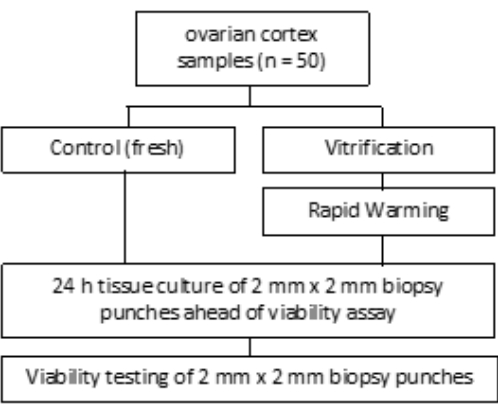
The overall success of the described vitrification protocol was assessed by comparing the vital follicle count before and after vitrification/rapid warming. The experimental setup is shown in Figure 1, and the results are presented in Figure 6. In 50 patients, a mean count of 77.98 vital follicles was observed before vitrification and 62.99 after vitrification/rapid warming, reflecting a survival rate of 80.8%. This was not significantly different according to the Wilcoxon test33.
Metal grids are individually customized with sharp scissors to a size of 25 mm x 8 mm, which fits into the caps of 1.8 mL cryovials, as shown in Figure 2A,C. After sterilization using steam autoclaving, the grids and caps of 1.8 mL vials are assembled under a laminar flow bench, as indicated in Figure 2B. This setup provides a secure hold in the cap without the need for additional actions and offers sufficient area for tissue of various sizes. Typically, tissue pieces of 5 mm x 10 mm for transplantation and 2 mm diameter pieces for assessing vital follicle count after rapid thawing are vitrified. Both sizes fit perfectly onto the metal grids.
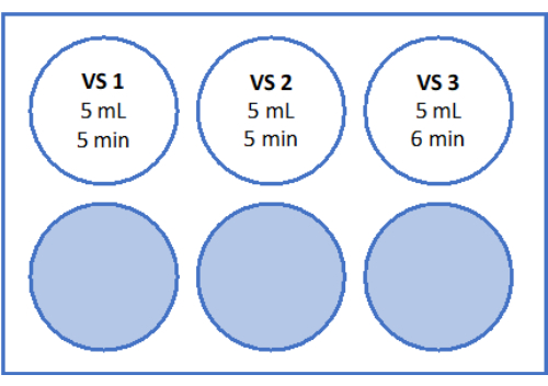
Equilibration with vitrification solutions (VS1, VS2, and VS3) is conducted in a 6-well plate on a rocking shaker at room temperature under a laminar flow bench, as shown in Figure 3. The time frames in the scheme ensure the effective uptake of the cryoprotectant ethylene glycol. Using a 6-well plate over single vessels is recommended as it facilitates quick movement of the tissue between solutions and helps prevent mix-ups.
For rapid vertical vitrification in liquid nitrogen, ovarian cortex tissue is cut into suitable pieces (Figure 4A). Ovarian cortex samples are placed on loading devices (Figure 4B,C) and submerged vertically into liquid nitrogen (Figure 4D) to achieve a glassy state of aggregation through vitrification. The chosen metal grids allow for vertical handling of the tissue, as shown in Figure 4D. Additionally, the metal grids are highly thermally conductive, ensuring rapid cooling rates from 22 °C to -196 °C, which is a critically important step in the vitrification process.
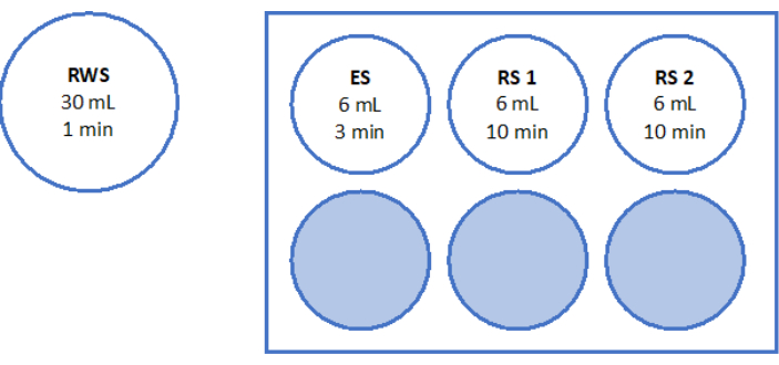
For rapid warming, RWS is prepared in a sterile cup at 37 °C. ES, RS1, and RS2 are prepared in a 6-well plate on a rocking shaker at room temperature, as shown in Figure 5. The high volume of pre-warmed RWS prevents the solution from cooling down excessively upon the addition of the vitrified tissue and ensures a consistently warm environment for the tissue throughout the rapid warming process.
To assess and ensure high quality standards, 2 mm x 2 mm biopsy punches are stained with calcein prior to vitrification and after rapid warming to determine follicular viability using fluorescence microscopy34,35 (Figure 6). Viable follicles emit green fluorescence at 495 nm after the intracellular uptake of calcein. Alternatively, follicular viability (Table 3) can be assessed using neutral red dyes36.
By following the described steps in this protocol, ovarian tissue is transformed into a glassy state of aggregation, which facilitates high survival rates after rapid warming, as confirmed by fluorescence microscopy.

Figure 1: Study design. Ovarian cortical samples from 50 patients were examined before (fresh) and after vitrification and rapid warming for the number of viable follicles. For each group, two tissue pieces of 2 mm diameter were cultured for 24 h before follicle count assessment. The tissue was digested with collagenase and stained with calcein to assess viability. The number of viable follicles was determined using a microscope. Please click here to view a larger version of this figure.

Figure 2: Preparation of loading devices. Metal grids were cut to a size of 8 mm x 25 mm (A). The customized metal grids were sterilized by autoclaving (B). The sterilized metal grids were then inserted into caps of 1.8 mL vials, ready for use (C). Please click here to view a larger version of this figure.

Figure 3: Preparation of vitrification solutions. Vitrification solutions (VS) were prepared and transferred to a 6-well plate. The wells show the volume of each solution and the individual incubation times used in the vitrification protocol. Please click here to view a larger version of this figure.

Figure 4: Ovarian cortex processing and vitrification. Ovarian cortex tissue was processed for cryopreservation by removing the medulla and cutting the tissue into 5 mm x 10 mm pieces (A). After incubation in the vitrification solutions shown in Figure 3, the tissue was loaded onto the vitrification loading device (B,C). For rapid vertical vitrification of ovarian cortex samples, the caps with the loaded tissue were quickly inserted into sterilized liquid nitrogen (D). Please click here to view a larger version of this figure.

Figure 5: Preparation of rapid warming solutions. Rapid warming solution (RWS), equilibration solution (ES), and rinsing solutions (RS) 1 and 2 were prepared and transferred to a 6-well plate. The vessels and wells show the volume of each solution and the corresponding incubation times. Note that RWS is maintained at 37.2 °C on a heating plate. Please click here to view a larger version of this figure.

Figure 6: Viability count. To assess the number of viable follicles, tissue pieces of 2 mm diameter were digested with collagenase and stained with calcein. The representative images show (A) calcein staining of 3 recovered follicles prior to vitrification. (B) A recovered viable follicle after vitrification and rapid warming. (C) A recovered viable follicle prior to slow freezing. (D) Recovered viable follicles after slow freezing and thawing. Follicular viability is indicated by calcein, a green fluorescent dye that emits green fluorescence when enzymatically converted by viable cells at 495 nm. Scale bar = 100 µm. Please click here to view a larger version of this figure.
| VS 1 (15 mL) | Ethylene glycol | 10% | 1.5 mL |
| SSS | 10% | 1.5 mL | |
| G-MOPS+ | 12 mL | ||
| VS 2 (15 mL) | Ethylene glycol | 20% | 3 mL |
| SSS | 10% | 1.5 mL | |
| G-MOPS+ | 10.5 mL | ||
| VS 3 (15 mL) | Ethylene glycol | 35% | 5.25 mL |
| SSS | 10% | 1.5 mL | |
| Sucrose | 0.5 mol/L | 2.57 g | |
| PVP | 5 % (w/v) | 0.75 g | |
| G-MOPS+ | ad 15 mL |
Table 1: Composition of vitrification solutions (VS).
| RWS (30 mL) | Sucrose | 0.8 mol/L | |
| 8.22 g | |||
| SSS | 10% | 3 mL | |
| G-MOPS+ | ad 30 mL | ||
| ES (15 mL) | Sucrose | 0.4 mol/L | 2.05 g |
| SSS | 10% | 1.5 mL | |
| G-MOPS+ | ad 15 mL | ||
| RS 1&2 (15 mL) | SSS | 10% | 1.5 mL |
| G-MOPS+ | ad 15 mL |
Table 2: Composition of rapid warming solution (RWS), equilibration solution (ES), and rinsing solutions (RS). This table provides the components and concentrations for the rapid warming solution (RWS), equilibration solution (ES), and rinsing solutions (RS) used in the post-vitrification processing.
| Parameter | Fresh | Interval | Rapid warmed after vitrification | Interval | n | *P-value |
| SD | SD | |||||
| Follicular viability count [n] | 77.98 | 0-386 | 62.99 | 0.5-349 | 50 | 0.130 |
| 77.95 | 80.02 | |||||
| *Wilcoxon test |
Table 3: Representative results of follicular viability. This table presents the results of follicular viability assessments before cryopreservation and after rapid warming. Two tissue pieces of 2 mm diameter per patient were used to count the number of viable follicles before and after vitrification/rapid warming. Paired tissue samples from 50 patients were analyzed using the Wilcoxon test.
Discussion
Here, a protocol for high-throughput vitrification of human ovarian cortex tissue, suitable for clinical routine, is presented. Similar to the vitrification of oocytes or embryos, the successful application of the procedure requires detailed adherence to the protocol concerning the temperature of the vitrification and warming solutions, as well as the equilibration period. Compliance with EU tissue directives37 regarding air quality and sterility is also essential.
The vitrification procedure results in a non-crystalline, amorphous, or glassy state. Overall, vitrification is a versatile process with significant implications in various scientific and technological domains. The primary benefit of vitrification is its ability to convert tissue into a glassy state, thereby preventing ice crystal formation38,39,40, which can negatively affect tissue integrity and its components.
Supplementation of cryoprotective agents (CPAs) with polyvinylpyrrolidone (PVP) allows for a reduction in CPA concentration without compromising the quality of the vitrification solutions41,42. Furthermore, the use of metal grids provides high thermal conductivity compared to plastic-based carrier systems. The grid structure also facilitates surface adhesion, ensuring the safe and secure cryopreservation of tissue samples and small cortex punches for quality measures. If cryovessels from other manufacturers are used, it is important to test the size of the metal grids beforehand to ensure stability and proper grip within the cryovessel lid, as well as a good fit into the vessel.
Critical steps to ensure successful vitrification include rapid vitrification by immersing the tissue in sterilized liquid nitrogen and performing rapid warming without delay to avoid adverse results. In terms of cost-effectiveness, tissue vitrification is less demanding compared to the slow freezing procedure, which may influence personnel deployment planning. Additionally, vitrification eliminates the need to purchase and service equipment required for slow freezing.
Biological measures and meta-analyses have demonstrated the comparability or even advantages of vitrification compared to slow freezing43. However, differences in results after vitrification may be attributed to the lack of standardization in both the vitrification device and the protocol, including the solutions used, which vary across studies. Future research should explore the potential for follicle culture from vitrified/rapid-warmed tissue to monitor growth in vitro, as successfully demonstrated in mouse ovarian tissue by several groups44,45,46,47,48,49,50.
In summary, vitrification of ovarian tissue is a significant alternative to the widely used slow freezing protocol, supported by five successful deliveries reported by Suzuki51 (Japan), Silber52 (USA), and Sänger53 (Germany). In contrast to commercially available vitrification media and kits for cells, there are few FDA/CE-approved systems for ovarian tissue, which may limit their application in clinical settings. Therefore, the development of FDA/CE-approved kits and media for the vitrification and rapid warming of ovarian tissue is recommended30.
Disclosures
None.
Acknowledgements
We thank Cara Färber for proofreading; Katharina Wollersheim, Martin Mahlberg, Lea Korte, and Jasmin Rebholz for their technical assistance.
Materials
| Name | Company | Catalog Number | Comments |
| 1.8 mL vials | VWR International GmbH | 479-6837 | |
| 10 mL serological pipette | Sarstedt | 86.1254.001 | |
| 4 well plate | Gynemed | GYOOPW-FW04 | |
| 50 mL Tube | Sarstedt | 62.559.001 | |
| 6 well plates | Sarstedt | 83.3920 | |
| Bacillol AF | Hartmann | 973385 | |
| Calcein AM | Merck | 17783 | |
| Collagenase type 1A | Merck | C2674 | |
| Cryosure DMSO | WAK Chemie | WAK-DMSO-10 | |
| Custodiol | Dr. Franz Köhler Chemie | 00867288 | |
| DPBS CTS | Gibco Life technologies | A12856-01 | |
| ErgoOne pipette aid | Starlab | S7166-0010 | |
| Ethylene glycol | Sigma Aldrich | 102466 | |
| Euronda sterilization container | euronda | 282021 | |
| G-MOPS+ | Vitrolife | 10130 | |
| Metal meshes | Sigma Aldrich | S0770 | |
| Metzenbaum scissors | world precision instruments | 501262102 | |
| N-Bath System | Nterilizer | N-Bath 3.0 | |
| Polyvinylpyrrolidone (PVP) | SAGE | ART-4005 | |
| Serum substitute supplement (SSS) | Fujifilm Irvine scientific | 99193 | |
| Sterile cup | Sarstedt | 75.562.105 | |
| Sterile forceps | Carl Roth | KL05.1 | |
| Sucrose | Merck | S0389 |
References
- Rezazadeh Valojerdi, M., Eftekhari-Yazdi, P., Karimian, L., Hassani, F., Movaghar, B. Vitrification versus slow freezing gives excellent survival, post-warming embryo morphology and pregnancy outcomes for human cleaved embryos. J Assist Reprod Genet. 26 (6), 347-354 (2009).
- Levi-Setti, P. E., Patrizio, P., Scaravelli, G. Evolution of human oocyte cryopreservation: Slow freezing versus vitrification. Curr Opin Endocrinol Diabetes Obes. 2 (6), 445-450 (2016).
- Glujovsky, D., et al. Vitrification versus slow freezing for women undergoing oocyte cryopreservation. Cochrane Database Syst Rev. 9, CD010047 (2014).
- AbdelHafez, F. F., Desai, N., Abou-Setta, A. M., Falcone, T., Goldfarb, J. Slow freezing, vitrification and ultra-rapid freezing of human embryos: A systematic review and meta-analysis. Reprod Biomed Online. 20 (2), 209-222 (2010).
- Amorim, C. A., Curaba, M., Van Langendonckt, A., Dolmans, M. M., Donnez, J. Vitrification as an alternative means of cryo-preserving ovarian tissue. Reprod Biomed Online. 23, 160-186 (2011).
- Fahy, G. M., MacFarlane, D. R., Angell, C. A., Meryman, H. T. Vitrification as an approach to cryopreservation. Cryobiology. 21, 407-426 (1984).
- Liebermann, J., et al. Potential importance of vitrification in reproductive medicine. Biol Reprod. 67 (6), 1671-1680 (2002).
- Schallmoser, A., et al. Comparison of angiogenic potential in vitrified vs. slow frozen human ovarian tissue. Sci Rep. 13 (1), 12885 (2023).
- Schallmoser, A., et al. The effect of high-throughput vitrification of human ovarian cortex tissue on follicular viability: A promising alternative to conventional slow freezing. Arch Gynecol Obstet. 307 (2), 591-599 (2023).
- Jadoul, P., et al. Efficacy of ovarian tissue cryopreservation for fertility preservation: Lessons learned from 545 cases. Hum Reprod. 32 (5), 1046-1054 (2017).
- Meirow, D., et al. Pregnancy after transplantation of cryopreserved ovarian tissue in a patient with ovarian failure after chemotherapy. N Engl J Med. 353, 318-321 (2005).
- Meirow, D., et al. Transplantations of frozen-thawed ovarian tissue demonstrate high reproductive performance and the need to revise restrictive criteria. Fertil Steril. 106, 467-474 (2016).
- Hoekman, E. J., et al. Ovarian tissue cryopreservation: Low usage rates and high live-birth rate after transplantation. Acta Obstet Gynecol Scand. 00, 1-9 (2019).
- Rodriguez-Wallberg, K. A., et al. 86 Successful births and 9 ongoing pregnancies worldwide in women transplanted with frozen-thawed ovarian tissue: Focus on birth and perinatal outcome in 40 of these children. J Assist Reprod Genet. 34, 325-336 (2017).
- Anderson, R. A., et al. The ESHRE guideline group on female fertility preservation, ESHRE guideline: Female fertility preservation. Hum Reprod Open. 2020 (4), hoaa052 (2020).
- Shi, Q., Xie, Y., Wang, Y., Li, S. Vitrification versus slow freezing for human ovarian tissue cryopreservation: a systematic review and meta-analysis. Sci Rep. 7 (1), 8538 (2017).
- Keros, V., et al. Vitrification versus controlled rate freezing in cryopreservation of human ovarian tissue. Hum Reprod. 24, 1670-1683 (2009).
- Xiao, Z., Wang, Y., Li, L., Luo, S., Li, S. W. Needle immersed vitrification can lower the concentration of cryoprotectant in human ovarian tissue cryopreservation. Fertil Steril. 94, 2323-2328 (2010).
- Fabbri, R., et al. Good preservation of stromal cells and no apoptosis in human ovarian tissue after vitrification. Biomed Res Int. 2014, 673537 (2014).
- Chang, H. J., et al. Optimal condition of vitrification method for cryopreservation of human ovarian cortical tissues. J Obstet Gynaecol Res. 37 (8), 1092-1101 (2011).
- Wang, Y., Xiao, Z., Li, L., Fan, W., Li, S. W. Novel needle immersed vitrification: A practical and convenient method with potential advantages in mouse and human ovarian tissue cryopreservation. Hum Reprod. 23 (10), 2256-2265 (2020).
- Fabbri, R., et al. Morphological, ultrastructural and functional imaging of frozen/thawed and vitrified/warmed human ovarian tissue retrieved from oncological patients. Hum Reprod. 31 (8), 1838-1849 (2023).
- Xiao, Z., Wang, Y., Li, L. L., Li, S. W. In vitro culture thawed human ovarian tissue: NIV versus slow freezing method. Cryo Letters. 34 (5), 520-526 (2013).
- Locatelli, Y., et al. In vitro survival of follicles in prepubertal ewe ovarian cortex cryopreserved by slow freezing or non-equilibrium vitrification. J Assist Reprod Genet. 36 (9), 1823-1835 (2017).
- Nikiforov, D., et al. Innovative multi-protectoral approach increases survival rate after vitrification of ovarian tissue and isolated follicles with improved results in comparison with conventional method. J Ovarian Res. 11 (1), 65 (2018).
- Wang, T., et al. Human single follicle growth in vitro from cryopreserved ovarian tissue after slow freezing or vitrification. Human Reprod. 31 (4), 763-773 (2016).
- Lee, S., et al. Comparison between slow freezing and vitrification for human ovarian tissue cryopreservation and xenotransplantation. Int JMol Sci. 20 (13), 3346 (2019).
- Suzuki, N., et al. Successful fertility preservation following ovarian tissue vitrification in patients with primary ovarian insufficiency. Hum Reprod. 30 (3), 608-615 (2015).
- Silber, S. J., et al. Cryopreservation and transplantation of ovarian tissue: Results from one center in the USA. J Assist Reprod Genet. 35 (12), 2205-2213 (2018).
- Sänger, N., John, J., Einenkel, R., Schallmoser, A. First report on successful delivery after retransplantation of vitrified, rapid warmed ovarian tissue in Europe. Reprod Biomed Online. 49 (1), 103940 (2024).
- Sugishita, Y., et al. Quantification of residual cryoprotectants and cytotoxicity in thawed bovine ovarian tissues after slow freezing or vitrification. Hum Reprod. 37 (3), 522-533 (2022).
- Abir, R., et al. Attempts to improve human ovarian transplantation outcomes of needle-immersed vitrification and slow-freezing by host and graft treatments. J Assist Reprod Genet. 34 (5), 633-644 (2017).
- Sänger, N., John, J., Einenkel, R., Schallmoser, A. First report on successful delivery after retransplantation of vitrified, rapid warmed ovarian tissue in Europe. Reprod Biomed. 49 (1), 103940 (2024).
- Schallmoser, A., Einenkel, R., Färber, C., Sänger, N. In vitro growth (IVG) of human ovarian follicles in frozen thawed ovarian cortex tissue culture supplemented with follicular fluid under hypoxic conditions. Arch Gynecol Obstet. 306 (4), 1299-1311 (2022).
- Kristensen, S. G., et al. A simple method to quantify follicle survival in cryopreserved human ovarian tissue. Hum Reprod. 33 (12), 2276-2284 (2018).
- Mortimer, D. A critical assessment of the impact of the European Union Tissues and Cells Directive (2004) on laboratory practices in assisted conception. Reprod Biomed. 11 (2), 162-176 (2005).
- Amorim, C. A., Curaba, M., Van Langendonckt, A., Dolmans, M. M., Donnez, J. Vitrification as an alternative means of cryopreserving ovarian tissue. Reprod Biomed. 23 (2), 160-186 (2011).
- Fahy, G. M., Meryman, H. T. Vitrification: A new approach to organ cryopreservation. Transplantation: Approaches to Graft Rejection. , 305-335 (1986).
- Kattera, S., Chen, C. Cryopreservation of embryos by vitrification: Current development. Int Surg. 91 (5 Suppl), S55-S62 (2006).
- Fuller, B., Paynter, S. Fundamentals of cryobiology in reproductive medicine. Reprod Biomed. 9, 680-691 (2004).
- Liebermann, J., et al. Potential importance of vitrification in reproductive medicine. Biol Reprod. 67, 1671-1680 (2002).
- Shi, Q., Xie, Y., Wang, Y., Li, S. Vitrification versus slow freezing for human ovarian tissue cryopreservation: a systematic review and meta-analysis. Sci Rep. 7 (1), 8538 (2017).
- Hasegawa, A., Hamada, Y., Mehandjiev, T., Koyama, K. In vitro growth and maturation as well as fertilization of mouse preantral oocytes from vitrified ovaries. Fertil Steril. 81 (Suppl 1), 824-830 (2004).
- Segino, M., et al. In vitro culture of mouse GV oocytes and preantral follicles isolated from ovarian tissues cryopreserved by vitrification. Hum Cell. 16 (3), 109-116 (2003).
- Kagawa, N., et al. Production of the first offspring from oocytes derived from fresh and cryopreserved pre-antral follicles of adult mice. Reprod Biomed. 14 (6), 693-699 (2007).
- Haidari, K., et al. The effects of different concentrations of leukemia inhibitory factor on the development of isolated preantral follicles from fresh and vitrified mouse ovaries. Iran Biomed J. 10, 4 (2006).
- Haidari, K., Salehnia, M., Rezazadeh Valojerdi, M. The effect of leukemia inhibitory factor and coculture on the in vitro maturation and ultrastructure of vitrified and nonvitrified isolated mouse preantral follicles. Fertil Steril. 90 (6), 2389-2397 (2008).
- Lin, T. C., et al. Comparison of the developmental potential of 2-week-old preantral follicles derived from vitrified ovarian tissue slices, vitrified whole ovaries and vitrified/transplanted newborn mouse ovaries using the metal surface method. BMC Biotechnol. 8, 38 (2008).
- Wang, X., Catt, S., Pangestu, M., Temple-Smith, P. Live offspring from vitrified blastocysts derived from fresh and cryopreserved ovarian tissue grafts of adult mice. Reproduction. 138 (3), 527-535 (2009).
- Suzuki, N., et al. Successful fertility preservation following ovarian tissue vitrification in patients with primary ovarian insufficiency. Hum Reprod. 30 (3), 608-615 (2015).
- Silber, S. J., et al. Cryopreservation and transplantation of ovarian tissue: Results from one center in the USA. J Assist Reprodu Genet. 35 (12), 2205-2213 (2018).
- Sänger, N., John, J., Einenkel, R., Schallmoser, A. First report on successful delivery after retransplantation of vitrified, rapid warmed ovarian tissue in Europe. Reprod Biomed. 49 (1), 103940 (2024).
- Parmegiani, L., et al. Testing the efficacy and efficiency of a single "universal warming protocol" for vitrified human embryos: prospective randomized controlled trial and retrospective longitudinal cohort study. J Assist Reprod Gen. 35 (10), 1887-1895 (2018).
Reprints and Permissions
Request permission to reuse the text or figures of this JoVE article
Request PermissionThis article has been published
Video Coming Soon
Copyright © 2025 MyJoVE Corporation. All rights reserved